 Movie
Movie Controller

[English] 日本語
 Yorodumi
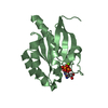
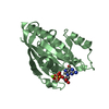
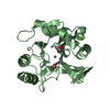
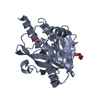
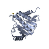
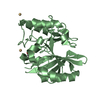
- PDB-1y43: crystal structure of aspergilloglutamic peptidase from Aspergillu... -
+ Open data

- Basic information
| Entry | Database: PDB / ID: 1y43 | ||||||
|---|---|---|---|---|---|---|---|
| Title | crystal structure of aspergilloglutamic peptidase from Aspergillus niger | ||||||
 Components Components |
| ||||||
 Keywords Keywords | HYDROLASE / aspergillopepsin II / proctase A / beta sandwich structure | ||||||
| Function / homology |  Function and homology information Function and homology informationaspergillopepsin II / glutamic-type endopeptidase activity / aspartic-type endopeptidase activity / proteolysis Similarity search - Function | ||||||
| Biological species |  | ||||||
| Method |  X-RAY DIFFRACTION / SYNCHROTRON / MIR / Resolution: 1.4 Å X-RAY DIFFRACTION / SYNCHROTRON / MIR / Resolution: 1.4 Å | ||||||
 Authors Authors | Sasaki, H. / Nakagawa, A. / Iwata, S. / Muramatsu, T. / Suganuma, M. / Sawano, Y. / Kojima, M. / Kubota, K. / Takahashi, K. | ||||||
 Citation Citation | Journal: Proc.Jpn.Acad.,Ser.B / Year: 2004 Title: The three-dimensional structure of aspergilloglutamic peptidase from Aspergillus niger Authors: Sasaki, H. / Nakagawa, A. / Muramatsu, T. / Suganuma, M. / Sawano, Y. / Kojima, M. / Kubota, K. / Takahashi, K. / Tanokura, M. | ||||||
| History |
|
- Structure visualization
| Structure viewer | Molecule: MolmilJmol/JSmol |
|---|
- Downloads & links
-Download
| PDBx/mmCIF format | 1y43.cif.gz | 48.6 KB | Display | PDBx/mmCIF format |
|---|---|---|---|---|
| PDB format | pdb1y43.ent.gz | 38.1 KB | Display | PDB format |
| PDBx/mmJSON format | 1y43.json.gz | Tree view | PDBx/mmJSON format | |
| Others |  Other downloads Other downloads |
-Validation report
| Summary document | 1y43_validation.pdf.gz | 376.1 KB | Display | wwPDB validaton report |
|---|---|---|---|---|
| Full document | 1y43_full_validation.pdf.gz | 376 KB | Display | |
| Data in XML | 1y43_validation.xml.gz | 5.3 KB | Display | |
| Data in CIF | 1y43_validation.cif.gz | 8.3 KB | Display | |
| Arichive directory | https://data.pdbj.org/pub/pdb/validation_reports/y4/1y43ftp://data.pdbj.org/pub/pdb/validation_reports/y4/1y43 | HTTPS FTP |
-Related structure data
| Similar structure data |
|---|


-Links
 PDBj
PDBj- Assembly
| Deposited unit | 
| ||||||||
|---|---|---|---|---|---|---|---|---|---|
| 1 |
| ||||||||
| Unit cell |
|
-Components
| #1: Protein/peptide | Mass: 3919.114 Da / Num. of mol.: 1 / Source method: isolated from a natural source / Source: (natural) | ||
|---|---|---|---|
| #2: Protein | Mass: 18372.299 Da / Num. of mol.: 1 / Source method: isolated from a natural source / Source: (natural) | ||
| #3: Chemical | ChemComp-SO4 /   Mass: 96.063 Da / Num. of mol.: 5 / Source method: obtained synthetically / Formula: SO4 Mass: 96.063 Da / Num. of mol.: 5 / Source method: obtained synthetically / Formula: SO4#4: Water | ChemComp-HOH / |  Mass: 18.015 Da / Num. of mol.: 128 / Source method: isolated from a natural source / Formula: H2O Mass: 18.015 Da / Num. of mol.: 128 / Source method: isolated from a natural source / Formula: H2O |
-Experimental details
-Experiment
| Experiment | Method: X-RAY DIFFRACTION / Number of used crystals: 1 |
|---|
- Sample preparation
| Crystal | Density Matthews: 1.64 Å3/Da / Density % sol: 26 % |
|---|---|
| Crystal grow | Temperature: 298 K / Method: vapor diffusion, hanging drop / pH: 2.1 Details: ammonium sulfate, dimethyl sulfoxide, glycine, pH 2.1, VAPOR DIFFUSION, HANGING DROP, temperature 298K |
-Data collection
| Diffraction | Mean temperature: 283 K |
|---|---|
| Diffraction source | Source: SYNCHROTRON / Site: Photon Factory  / Beamline: BL-6A / Wavelength: 1 Å / Beamline: BL-6A / Wavelength: 1 Å |
| Detector | Type: FUJI / Detector: IMAGE PLATE / Date: Nov 30, 1993 |
| Radiation | Monochromator: Si / Protocol: SINGLE WAVELENGTH / Monochromatic (M) / Laue (L): M / Scattering type: x-ray |
| Radiation wavelength | Wavelength: 1 Å / Relative weight: 1 |
| Reflection | Resolution: 1.4→100 Å / Num. obs: 29424 / % possible obs: 97.5 % / Observed criterion σ(F): 1 / Observed criterion σ(I): 1 |
| Reflection shell | Resolution: 1.4→1.44 Å / % possible all: 82.5 |
- Processing
| Software |
| ||||||||||||||||||||
|---|---|---|---|---|---|---|---|---|---|---|---|---|---|---|---|---|---|---|---|---|---|
| Refinement | Method to determine structure: MIR / Resolution: 1.4→10 Å / σ(F): 1 / Stereochemistry target values: Engh & Huber
| ||||||||||||||||||||
| Refinement step | Cycle: LAST / Resolution: 1.4→10 Å
| ||||||||||||||||||||
| LS refinement shell | Resolution: 1.4→1.46 Å
|
