 Movie
Movie Controller

[English] 日本語
 Yorodumi
- PDB-1xxs: Structural insights for fatty acid binding in a Lys49 phospholipa... -
+ Open data

- Basic information
| Entry | Database: PDB / ID: 1xxs | ||||||
|---|---|---|---|---|---|---|---|
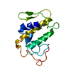
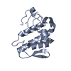
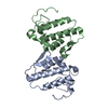
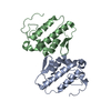
| Title | Structural insights for fatty acid binding in a Lys49 phospholipase A2: crystal structure of myotoxin II from Bothrops moojeni complexed with stearic acid | ||||||
 Components Components | Phospholipase A2 homolog 2 | ||||||
 Keywords Keywords | HYDROLASE / phospholipase A2 / stearic acid / dimer interface / fatty acid binding | ||||||
| Function / homology |  Function and homology information Function and homology information: / arachidonate secretion / phospholipid metabolic process / defense response to fungus / negative regulation of T cell proliferation / lipid catabolic process / phospholipid binding / toxin activity / killing of cells of another organism / defense response to bacterium ...: / arachidonate secretion / phospholipid metabolic process / defense response to fungus / negative regulation of T cell proliferation / lipid catabolic process / phospholipid binding / toxin activity / killing of cells of another organism / defense response to bacterium / calcium ion binding / extracellular region Similarity search - Function | ||||||
| Biological species |  Bothrops moojeni (Brazilian lancehead) Bothrops moojeni (Brazilian lancehead) | ||||||
| Method |  X-RAY DIFFRACTION / SYNCHROTRON / MOLECULAR REPLACEMENT / Resolution: 1.8 Å X-RAY DIFFRACTION / SYNCHROTRON / MOLECULAR REPLACEMENT / Resolution: 1.8 Å | ||||||
 Authors Authors | Watanabe, L. / Soares, A.M. / Ward, R.J. / Fontes, M.R. / Arni, R.K. | ||||||
 Citation Citation | Journal: Biochimie / Year: 2005 Title: Structural insights for fatty acid binding in a Lys49-phospholipase A(2): crystal structure of myotoxin II from Bothrops moojeni complexed with stearic acid Authors: Watanabe, L. / Soares, A.M. / Ward, R.J. / Fontes, M.R. / Arni, R.K. #1: Journal: PROTEIN PEPT.LETT. / Year: 2003Title: Initiating structural studies of Lys49-PLA2 homologues complexed with an anionic detergent, a fatty acid and a natural lipid Authors: Watanabe, L. / Fontes, M.R. / Soares, A.M. / Giglio, J.R. / Arni, R.K. #2: Journal: Arch.Biochem.Biophys. / Year: 2000Title: Structural and functional characterization of myotoxin I, a Lys49 phospholipase A(2) homologue from Bothrops moojeni (Caissaca) snake venom Authors: Soares, A.M. / Andriao-Escarso, S.H. / Angulo, Y. / Lomonte, B. / Gutierrez, J.M. / Marangoni, S. / Toyama, M.H. / Arni, R.K. / Giglio, J.R. #3: Journal: Toxicon / Year: 1998Title: A rapid procedure for the isolation of the Lys-49 myotoxin II from Bothrops moojeni (caissaca) venom: biochemical characterization, crystallization, myotoxic and edematogenic activity Authors: Soares, A.M. / Rodrigues, V.M. / Homsi-Brandeburgo, M.I. / Toyama, M.H. / Lombardi, F.R. / Arni, R.K. / Giglio, J.R. #4: Journal: PROTEIN PEPT.LETT. / Year: 1997Title: Crystal Structure of miotoxin-II: a myotoxic phospholipase A2 homologue from Bothrops moojeni venom Authors: de Azevedo Jr., W.F. / Ward, R.J. / Lombardi, F.R. / Giglio, J.R. / Soares, A.M. / Fontes, M.R.M. / Arni, R.K. | ||||||
| History |
| ||||||
| Remark 600 | HETEROGEN STE 205 AND STE 206 ARE BOUND AT THE DIMER INTERFACE OF THE PROTEIN. THEY ARE RELATED BY ...HETEROGEN STE 205 AND STE 206 ARE BOUND AT THE DIMER INTERFACE OF THE PROTEIN. THEY ARE RELATED BY TWO-FOLD SYMMETRY AND SHARE THE SAME SITE. THESE MOLECULES WERE REFINED WITH PARTIAL OCCUPANCIES (0.5). |
- Structure visualization
| Structure viewer | Molecule: MolmilJmol/JSmol |
|---|
- Downloads & links
-Download
| PDBx/mmCIF format | 1xxs.cif.gz | 63.9 KB | Display | PDBx/mmCIF format |
|---|---|---|---|---|
| PDB format | pdb1xxs.ent.gz | 47.8 KB | Display | PDB format |
| PDBx/mmJSON format | 1xxs.json.gz | Tree view | PDBx/mmJSON format | |
| Others |  Other downloads Other downloads |
-Validation report
| Arichive directory | https://data.pdbj.org/pub/pdb/validation_reports/xx/1xxsftp://data.pdbj.org/pub/pdb/validation_reports/xx/1xxs | HTTPS FTP |
|---|
-Related structure data
| Related structure data | |
|---|---|
| Similar structure data |


-Links
 PDBj
PDBj

- Assembly
| Deposited unit | 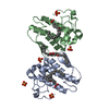
| ||||||||
|---|---|---|---|---|---|---|---|---|---|
| 1 |
| ||||||||
| Unit cell |
|
-Components
| #1: Protein | Mass: 13836.114 Da / Num. of mol.: 2 / Source method: isolated from a natural source / Details: venom glands / Source: (natural) Bothrops moojeni (Brazilian lancehead) / References: UniProt: Q9I834, phospholipase A2#2: Chemical | ChemComp-SO4 /   Mass: 96.063 Da / Num. of mol.: 5 / Source method: obtained synthetically / Formula: SO4 Mass: 96.063 Da / Num. of mol.: 5 / Source method: obtained synthetically / Formula: SO4#3: Chemical | ChemComp-STE /   Mass: 284.477 Da / Num. of mol.: 6 / Source method: obtained synthetically / Formula: C18H36O2 Mass: 284.477 Da / Num. of mol.: 6 / Source method: obtained synthetically / Formula: C18H36O2Has protein modification | Y | |
|---|
-Experimental details
-Experiment
| Experiment | Method: X-RAY DIFFRACTION / Number of used crystals: 1 |
|---|
- Sample preparation
| Crystal | Density Matthews: 2.58 Å3/Da / Density % sol: 52.03 % |
|---|---|
| Crystal grow | Temperature: 291 K / Method: vapor diffusion, hanging drop / pH: 6 Details: 0.6-0.8 M sodium citrate, pH 6.0, VAPOR DIFFUSION, HANGING DROP, temperature 291K |
-Data collection
| Diffraction | Mean temperature: 298 K |
|---|---|
| Diffraction source | Source: SYNCHROTRON / Site: LNLS  / Beamline: D03B-MX1 / Wavelength: 1.38 Å / Beamline: D03B-MX1 / Wavelength: 1.38 Å |
| Detector | Type: MARRESEARCH / Detector: IMAGE PLATE / Date: Apr 15, 1999 |
| Radiation | Monochromator: mirrors / Protocol: SINGLE WAVELENGTH / Monochromatic (M) / Laue (L): M / Scattering type: x-ray |
| Radiation wavelength | Wavelength: 1.38 Å / Relative weight: 1 |
| Reflection | Resolution: 1.8→15.4 Å / Num. all: 21341 / Num. obs: 21341 / % possible obs: 80.7 % / Observed criterion σ(F): 0 / Observed criterion σ(I): 0 / Biso Wilson estimate: 27.3 Å2 / Rmerge(I) obs: 0.077 |
| Reflection shell | Resolution: 1.8→1.91 Å / Rmerge(I) obs: 0.221 / Num. unique all: 2787 / % possible all: 66.9 |
- Processing
| Software |
| |||||||||||||||||||||||||
|---|---|---|---|---|---|---|---|---|---|---|---|---|---|---|---|---|---|---|---|---|---|---|---|---|---|---|
| Refinement | Method to determine structure: MOLECULAR REPLACEMENT Starting model: MjTX-II native Resolution: 1.8→15.4 Å / Isotropic thermal model: Isotropic / Cross valid method: THROUGHOUT / σ(F): 0 / Stereochemistry target values: Engh & Huber
| |||||||||||||||||||||||||
| Displacement parameters | Biso mean: 35.5 Å2 | |||||||||||||||||||||||||
| Refine analyze | Luzzati coordinate error obs: 0.23 Å / Luzzati d res low obs: 4 Å / Luzzati sigma a obs: 0.3 Å | |||||||||||||||||||||||||
| Refinement step | Cycle: LAST / Resolution: 1.8→15.4 Å
| |||||||||||||||||||||||||
| Refine LS restraints |
|
