 Movie
Movie Controller


+ Open data

- Basic information
| Entry | Database: PDB / ID: 5kmx | ||||||
|---|---|---|---|---|---|---|---|
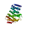
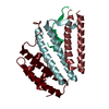
| Title | Structure of Trypanosoma congolense Insect Stage Antigen | ||||||
 Components Components | (Putative uncharacterized protein TCIL3000_10_9440) x 2 | ||||||
 Keywords Keywords | MEMBRANE PROTEIN / Bi-lobed architecture / surface protein / putative sensor | ||||||
| Function / homology | membrane / Uncharacterized protein TCIL3000_10_9440 Function and homology information Function and homology information | ||||||
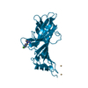
| Biological species |  | ||||||
| Method |  X-RAY DIFFRACTION / SYNCHROTRON / MOLECULAR REPLACEMENT / Resolution: 2.452 Å X-RAY DIFFRACTION / SYNCHROTRON / MOLECULAR REPLACEMENT / Resolution: 2.452 Å | ||||||
| Model details | Bi-lobed protein | ||||||
 Authors Authors | Ramaswamy, R. / Parker, M.L. / Boulanger, M.J. | ||||||
| Funding support |  Canada, 1items Canada, 1items
| ||||||
 Citation Citation | Journal: Protein Sci. / Year: 2016 Title: Structural characterization reveals a novel bilobed architecture for the ectodomains of insect stage expressed Trypanosoma brucei PSSA-2 and Trypanosoma congolense ISA. Authors: Ramaswamy, R. / Goomeshi Nobary, S. / Eyford, B.A. / Pearson, T.W. / Boulanger, M.J. | ||||||
| History |
|
- Structure visualization
| Structure viewer | Molecule: MolmilJmol/JSmol |
|---|
- Downloads & links
-Download
| PDBx/mmCIF format | 5kmx.cif.gz | 171.6 KB | Display | PDBx/mmCIF format |
|---|---|---|---|---|
| PDB format | pdb5kmx.ent.gz | 135.1 KB | Display | PDB format |
| PDBx/mmJSON format | 5kmx.json.gz | Tree view | PDBx/mmJSON format | |
| Others |  Other downloads Other downloads |
-Validation report
| Arichive directory | https://data.pdbj.org/pub/pdb/validation_reports/km/5kmxftp://data.pdbj.org/pub/pdb/validation_reports/km/5kmx | HTTPS FTP |
|---|
-Related structure data
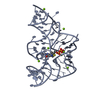
| Related structure data |  5klhSC S: Starting model for refinement C: citing same article ( |
|---|---|
| Similar structure data |


-Links
 PDBj
PDBj- Assembly
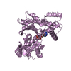
| Deposited unit | 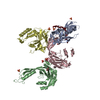
| ||||||||
|---|---|---|---|---|---|---|---|---|---|
| 1 | 
| ||||||||
| 2 | 
| ||||||||
| 3 | 
| ||||||||
| 4 | 
| ||||||||
| Unit cell |
| ||||||||
| Details | Monomer as determined by size exclusion chromatography. |
-Components
| #1: Protein | Mass: 26145.184 Da / Num. of mol.: 4 / Fragment: residues 40-264 Source method: isolated from a genetically manipulated source Source: (gene. exp.)  #2: Protein/peptide | | Mass: 1209.482 Da / Num. of mol.: 1 Source method: isolated from a genetically manipulated source Source: (gene. exp.) #3: Chemical | ChemComp-SO4 /   Mass: 96.063 Da / Num. of mol.: 8 / Source method: obtained synthetically / Formula: SO4 Mass: 96.063 Da / Num. of mol.: 8 / Source method: obtained synthetically / Formula: SO4#4: Chemical |   Mass: 92.094 Da / Num. of mol.: 2 / Source method: obtained synthetically / Formula: C3H8O3 Mass: 92.094 Da / Num. of mol.: 2 / Source method: obtained synthetically / Formula: C3H8O3#5: Water | ChemComp-HOH / |  Mass: 18.015 Da / Num. of mol.: 68 / Source method: isolated from a natural source / Formula: H2O Mass: 18.015 Da / Num. of mol.: 68 / Source method: isolated from a natural source / Formula: H2OHas protein modification | Y | |
|---|
-Experimental details
-Experiment
| Experiment | Method: X-RAY DIFFRACTION / Number of used crystals: 1 |
|---|
- Sample preparation
| Crystal | Density Matthews: 2.85 Å3/Da / Density % sol: 56.89 % |
|---|---|
| Crystal grow | Temperature: 291 K / Method: vapor diffusion / pH: 6.5 / Details: 20% PEG 3350 with 0.2 M (NH4)2SO4 |
-Data collection
| Diffraction | Mean temperature: 100 K |
|---|---|
| Diffraction source | Source: SYNCHROTRON / Site: SSRL  / Beamline: BL9-2 / Wavelength: 0.9795 Å / Beamline: BL9-2 / Wavelength: 0.9795 Å |
| Detector | Type: DECTRIS PILATUS 6M / Detector: PIXEL / Date: Nov 12, 2014 / Details: Rh coated flat, toroidal focusing |
| Radiation | Monochromator: Liquid nitrogen-cooled double crystal monochromator, non fixed exit slit Protocol: SINGLE WAVELENGTH / Monochromatic (M) / Laue (L): M / Scattering type: x-ray |
| Radiation wavelength | Wavelength: 0.9795 Å / Relative weight: 1 |
| Reflection | Resolution: 2.45→39.472 Å / Num. obs: 42706 / % possible obs: 97.6 % / Redundancy: 3.4 % / Biso Wilson estimate: 64.65 Å2 / CC1/2: 0.96 / Rmerge(I) obs: 0.044 / Net I/σ(I): 17.8 |
| Reflection shell | Resolution: 2.452→2.54 Å / Redundancy: 3.5 % / Rmerge(I) obs: 0.517 / Mean I/σ(I) obs: 1.2 / CC1/2: 0.821 / % possible all: 98.34 |
- Processing
| Software |
| ||||||||||||||||||||||||||||||||||||||||||||||||||||||||||||||||||||||||||||||||||||||||||||||||||||||||||||||||
|---|---|---|---|---|---|---|---|---|---|---|---|---|---|---|---|---|---|---|---|---|---|---|---|---|---|---|---|---|---|---|---|---|---|---|---|---|---|---|---|---|---|---|---|---|---|---|---|---|---|---|---|---|---|---|---|---|---|---|---|---|---|---|---|---|---|---|---|---|---|---|---|---|---|---|---|---|---|---|---|---|---|---|---|---|---|---|---|---|---|---|---|---|---|---|---|---|---|---|---|---|---|---|---|---|---|---|---|---|---|---|---|---|---|
| Refinement | Method to determine structure: MOLECULAR REPLACEMENT Starting model: 5KLH Resolution: 2.452→39.472 Å / SU ML: 0.44 / Cross valid method: FREE R-VALUE / σ(F): 1.37 / Phase error: 34.26
| ||||||||||||||||||||||||||||||||||||||||||||||||||||||||||||||||||||||||||||||||||||||||||||||||||||||||||||||||
| Solvent computation | Shrinkage radii: 0.9 Å / VDW probe radii: 1.11 Å | ||||||||||||||||||||||||||||||||||||||||||||||||||||||||||||||||||||||||||||||||||||||||||||||||||||||||||||||||
| Displacement parameters | Biso max: 156.95 Å2 / Biso mean: 64.65 Å2 / Biso min: 30 Å2 | ||||||||||||||||||||||||||||||||||||||||||||||||||||||||||||||||||||||||||||||||||||||||||||||||||||||||||||||||
| Refinement step | Cycle: final / Resolution: 2.452→39.472 Å
| ||||||||||||||||||||||||||||||||||||||||||||||||||||||||||||||||||||||||||||||||||||||||||||||||||||||||||||||||
| Refine LS restraints |
| ||||||||||||||||||||||||||||||||||||||||||||||||||||||||||||||||||||||||||||||||||||||||||||||||||||||||||||||||
| LS refinement shell | Refine-ID: X-RAY DIFFRACTION / Total num. of bins used: 15
|
