 Movie
Movie Controller

[English] 日本語
 Yorodumi
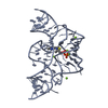
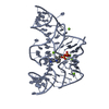
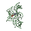
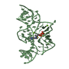
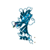
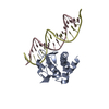
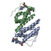
- PDB-3d2x: Structure of the thiamine pyrophosphate-specific riboswitch bound... -
+ Open data

- Basic information
| Entry | Database: PDB / ID: 3d2x | ||||||
|---|---|---|---|---|---|---|---|
| Title | Structure of the thiamine pyrophosphate-specific riboswitch bound to oxythiamine pyrophosphate | ||||||
 Components Components | TPP-specific riboswitch | ||||||
 Keywords Keywords | RNA / riboswitch / thiamine pyrophosphate / oxythiamine pyrophosphate / antibiotic / drugs / complex | ||||||
| Function / homology | Chem-D2X / RNA / RNA (> 10) Function and homology information Function and homology information | ||||||
| Biological species |  | ||||||
| Method |  X-RAY DIFFRACTION / SYNCHROTRON / MOLECULAR REPLACEMENT / Resolution: 2.5 Å X-RAY DIFFRACTION / SYNCHROTRON / MOLECULAR REPLACEMENT / Resolution: 2.5 Å | ||||||
 Authors Authors | Thore, S. / Frick, C. / Ban, N. | ||||||
 Citation Citation | Journal: J.Am.Chem.Soc. / Year: 2008 Title: Structural basis of thiamine pyrophosphate analogues binding to the eukaryotic riboswitch Authors: Thore, S. / Frick, C. / Ban, N. | ||||||
| History |
|
- Structure visualization
| Structure viewer | Molecule: MolmilJmol/JSmol |
|---|
- Downloads & links
-Download
| PDBx/mmCIF format | 3d2x.cif.gz | 99.3 KB | Display | PDBx/mmCIF format |
|---|---|---|---|---|
| PDB format | pdb3d2x.ent.gz | 74.1 KB | Display | PDB format |
| PDBx/mmJSON format | 3d2x.json.gz | Tree view | PDBx/mmJSON format | |
| Others |  Other downloads Other downloads |
-Validation report
| Arichive directory | https://data.pdbj.org/pub/pdb/validation_reports/d2/3d2xftp://data.pdbj.org/pub/pdb/validation_reports/d2/3d2x | HTTPS FTP |
|---|
-Related structure data
| Related structure data |  3d2gC  3d2vC  2ckyS S: Starting model for refinement C: citing same article ( |
|---|---|
| Similar structure data |


-Links
 PDBj
PDBj


- Assembly
| Deposited unit | 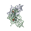
| ||||||||
|---|---|---|---|---|---|---|---|---|---|
| 1 | 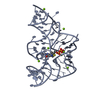
| ||||||||
| 2 | 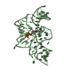
| ||||||||
| Unit cell |
|
-Components
| #1: RNA chain | Mass: 24958.838 Da / Num. of mol.: 2 Source method: isolated from a genetically manipulated source Source: (gene. exp.) #2: Chemical | ChemComp-MG /   Mass: 24.305 Da / Num. of mol.: 13 / Source method: obtained synthetically / Formula: Mg Mass: 24.305 Da / Num. of mol.: 13 / Source method: obtained synthetically / Formula: Mg#3: Chemical | 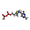  Mass: 426.299 Da / Num. of mol.: 2 / Source method: obtained synthetically / Formula: C12H18N3O8P2S Mass: 426.299 Da / Num. of mol.: 2 / Source method: obtained synthetically / Formula: C12H18N3O8P2S#4: Water | ChemComp-HOH / |  Mass: 18.015 Da / Num. of mol.: 126 / Source method: isolated from a natural source / Formula: H2O Mass: 18.015 Da / Num. of mol.: 126 / Source method: isolated from a natural source / Formula: H2O |
|---|
-Experimental details
-Experiment
| Experiment | Method: X-RAY DIFFRACTION / Number of used crystals: 1 |
|---|
- Sample preparation
| Crystal | Density Matthews: 2.33 Å3/Da / Density % sol: 47.16 % | ||||||||||||||||||||||||||||||||
|---|---|---|---|---|---|---|---|---|---|---|---|---|---|---|---|---|---|---|---|---|---|---|---|---|---|---|---|---|---|---|---|---|---|
| Crystal grow | Temperature: 298 K / Method: vapor diffusion, hanging drop / pH: 6.8 Details: 12-18% 1,6-hexanediol, 0.5mM spermine, 10mM magnesium sulfate, 40mM sodium cacodylate, pH 6.8, VAPOR DIFFUSION, HANGING DROP, temperature 298K | ||||||||||||||||||||||||||||||||
| Components of the solutions |
|
-Data collection
| Diffraction | Mean temperature: 100 K |
|---|---|
| Diffraction source | Source: SYNCHROTRON / Site: SLS  / Beamline: X06SA / Wavelength: 1 Å / Beamline: X06SA / Wavelength: 1 Å |
| Detector | Type: MARMOSAIC 225 mm CCD / Detector: CCD / Date: Aug 11, 2006 |
| Radiation | Monochromator: GRAPHITE / Protocol: SINGLE WAVELENGTH / Monochromatic (M) / Laue (L): M / Scattering type: x-ray |
| Radiation wavelength | Wavelength: 1 Å / Relative weight: 1 |
| Reflection | Resolution: 2.5→15 Å / Num. all: 16736 / Num. obs: 16268 / % possible obs: 97.2 % / Observed criterion σ(F): -3 / Observed criterion σ(I): -3 / Redundancy: 7.3 % / Biso Wilson estimate: 42.3 Å2 / Rmerge(I) obs: 0.153 / Net I/σ(I): 14.6 |
| Reflection shell | Resolution: 2.5→2.55 Å / Redundancy: 7.5 % / Rmerge(I) obs: 0.533 / Mean I/σ(I) obs: 2.85 / Num. unique all: 934 / % possible all: 97.1 |
- Processing
| Software |
| |||||||||||||||||||||||||
|---|---|---|---|---|---|---|---|---|---|---|---|---|---|---|---|---|---|---|---|---|---|---|---|---|---|---|
| Refinement | Method to determine structure: MOLECULAR REPLACEMENT Starting model: PDB entry 2cky Resolution: 2.5→15 Å / σ(F): 0 / Stereochemistry target values: Engh & Huber
| |||||||||||||||||||||||||
| Refine analyze | Luzzati coordinate error obs: 0.37 Å | |||||||||||||||||||||||||
| Refinement step | Cycle: LAST / Resolution: 2.5→15 Å
|
