 Movie
Movie Controller

[English] 日本語
 Yorodumi
- PDB-3t0r: Crystal Structure of MjTX-I, a myotoxic Lys49-phospholipase A2 fr... -
+ Open data

- Basic information
| Entry | Database: PDB / ID: 3t0r | ||||||
|---|---|---|---|---|---|---|---|
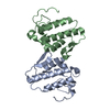
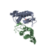
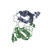
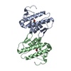
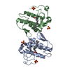
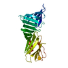
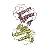
| Title | Crystal Structure of MjTX-I, a myotoxic Lys49-phospholipase A2 from Bothrops moojeni | ||||||
 Components Components | Phospholipase A2 homolog 1 | ||||||
 Keywords Keywords | TOXIN / Lys49-phospholipase A2 / Myotoxin / Venom Glands | ||||||
| Function / homology |  Function and homology information Function and homology informationA2-type glycerophospholipase activity / arachidonate secretion / phospholipid metabolic process / negative regulation of T cell proliferation / lipid catabolic process / phospholipid binding / toxin activity / calcium ion binding / extracellular region Similarity search - Function | ||||||
| Biological species |  Bothrops moojeni (Brazilian lancehead) Bothrops moojeni (Brazilian lancehead) | ||||||
| Method |  X-RAY DIFFRACTION / SYNCHROTRON / MOLECULAR REPLACEMENT / Resolution: 2.49 Å X-RAY DIFFRACTION / SYNCHROTRON / MOLECULAR REPLACEMENT / Resolution: 2.49 Å | ||||||
 Authors Authors | Salvador, G.H.M. / Marchi-Salvador, D.P. / Fontes, M.R.M. | ||||||
 Citation Citation | Journal: To be Published Title: Crystal Structure of MjTX-I, a myotoxic Lys49-phospholipase A2 from Bothrops moojeni Authors: Salvador, G.H.M. / Marchi-Salvador, D.P. / Fontes, M.R.M. | ||||||
| History |
|
- Structure visualization
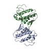
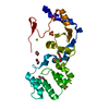
| Structure viewer | Molecule: MolmilJmol/JSmol |
|---|
- Downloads & links
-Download
| PDBx/mmCIF format | 3t0r.cif.gz | 190 KB | Display | PDBx/mmCIF format |
|---|---|---|---|---|
| PDB format | pdb3t0r.ent.gz | 152.6 KB | Display | PDB format |
| PDBx/mmJSON format | 3t0r.json.gz | Tree view | PDBx/mmJSON format | |
| Others |  Other downloads Other downloads |
-Validation report
| Arichive directory | https://data.pdbj.org/pub/pdb/validation_reports/t0/3t0rftp://data.pdbj.org/pub/pdb/validation_reports/t0/3t0r | HTTPS FTP |
|---|
-Related structure data
| Related structure data |  1qllS S: Starting model for refinement |
|---|---|
| Similar structure data |


-Links
 PDBj
PDBj
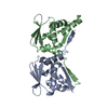
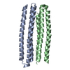
- Assembly
| Deposited unit | 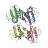
| ||||||||
|---|---|---|---|---|---|---|---|---|---|
| 1 | 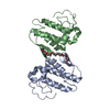
| ||||||||
| 2 | 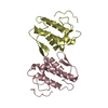
| ||||||||
| 3 | 
| ||||||||
| 4 | 
| ||||||||
| 5 | 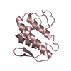
| ||||||||
| 6 | 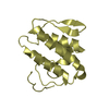
| ||||||||
| Unit cell |
| ||||||||
| Details | According to Small Angle X-ray Scattering experiments |
-Components
| #1: Protein | Mass: 13764.075 Da / Num. of mol.: 4 / Source method: isolated from a natural source / Source: (natural) Bothrops moojeni (Brazilian lancehead) / References: UniProt: P82114, phospholipase A2#2: Chemical | ChemComp-PE4 / |   Mass: 354.436 Da / Num. of mol.: 1 / Source method: obtained synthetically / Formula: C16H34O8 / Comment: precipitant*YM Mass: 354.436 Da / Num. of mol.: 1 / Source method: obtained synthetically / Formula: C16H34O8 / Comment: precipitant*YM#3: Water | ChemComp-HOH / |  Mass: 18.015 Da / Num. of mol.: 113 / Source method: isolated from a natural source / Formula: H2O Mass: 18.015 Da / Num. of mol.: 113 / Source method: isolated from a natural source / Formula: H2OHas protein modification | Y | |
|---|
-Experimental details
-Experiment
| Experiment | Method: X-RAY DIFFRACTION / Number of used crystals: 1 |
|---|
- Sample preparation
| Crystal | Density Matthews: 2.07 Å3/Da / Density % sol: 40.51 % |
|---|---|
| Crystal grow | Temperature: 291 K / Method: vapor diffusion, hanging drop Details: 32% (w/v) PEG 4000; 0.1 M Tris HCl; 0.15 M Magnesium chloride, pH 8.5, VAPOR DIFFUSION, HANGING DROP, temperature 291K PH range: 8,5 |
-Data collection
| Diffraction | Mean temperature: 100 K |
|---|---|
| Diffraction source | Source: SYNCHROTRON / Site: LNLS  / Beamline: D03B-MX1 / Wavelength: 1.421 Å / Beamline: D03B-MX1 / Wavelength: 1.421 Å |
| Detector | Type: MAR CCD 165 mm / Detector: CCD / Date: Aug 20, 2010 / Details: Mirrors |
| Radiation | Monochromator: Si curved crystal asymmetrically-cut / Protocol: SINGLE WAVELENGTH / Monochromatic (M) / Laue (L): M / Scattering type: x-ray |
| Radiation wavelength | Wavelength: 1.421 Å / Relative weight: 1 |
| Reflection | Resolution: 2.49→33.44 Å / Num. all: 15300 / Num. obs: 15300 / % possible obs: 98 % / Observed criterion σ(I): -3 / Redundancy: 3.1 % / Biso Wilson estimate: 43.016 Å2 / Rmerge(I) obs: 0.057 / Net I/σ(I): 20.64 |
| Reflection shell | Resolution: 2.49→2.55 Å / Redundancy: 3.1 % / Rmerge(I) obs: 0.22 / Mean I/σ(I) obs: 4.69 / Num. unique all: 1541 / % possible all: 98.9 |
- Processing
| Software |
| |||||||||||||||||||||||||||||||||||||||||||||||||||||||||||||||||||||||||||||||||||||||||||||||||||||||||||||||||||||||||||||
|---|---|---|---|---|---|---|---|---|---|---|---|---|---|---|---|---|---|---|---|---|---|---|---|---|---|---|---|---|---|---|---|---|---|---|---|---|---|---|---|---|---|---|---|---|---|---|---|---|---|---|---|---|---|---|---|---|---|---|---|---|---|---|---|---|---|---|---|---|---|---|---|---|---|---|---|---|---|---|---|---|---|---|---|---|---|---|---|---|---|---|---|---|---|---|---|---|---|---|---|---|---|---|---|---|---|---|---|---|---|---|---|---|---|---|---|---|---|---|---|---|---|---|---|---|---|---|
| Refinement | Method to determine structure: MOLECULAR REPLACEMENT Starting model: PDB ENTRY 1QLL Resolution: 2.49→33.44 Å / Cor.coef. Fo:Fc: 0.921 / Cor.coef. Fo:Fc free: 0.897 / SU B: 26.875 / SU ML: 0.267 / Cross valid method: THROUGHOUT / ESU R Free: 0.343 / Stereochemistry target values: MAXIMUM LIKELIHOOD / Details: HYDROGENS HAVE BEEN ADDED IN THE RIDING POSITIONS
| |||||||||||||||||||||||||||||||||||||||||||||||||||||||||||||||||||||||||||||||||||||||||||||||||||||||||||||||||||||||||||||
| Solvent computation | Ion probe radii: 0.8 Å / Shrinkage radii: 0.8 Å / VDW probe radii: 1.4 Å / Solvent model: BABINET MODEL WITH MASK | |||||||||||||||||||||||||||||||||||||||||||||||||||||||||||||||||||||||||||||||||||||||||||||||||||||||||||||||||||||||||||||
| Displacement parameters | Biso mean: 43.016 Å2
| |||||||||||||||||||||||||||||||||||||||||||||||||||||||||||||||||||||||||||||||||||||||||||||||||||||||||||||||||||||||||||||
| Refinement step | Cycle: LAST / Resolution: 2.49→33.44 Å
| |||||||||||||||||||||||||||||||||||||||||||||||||||||||||||||||||||||||||||||||||||||||||||||||||||||||||||||||||||||||||||||
| Refine LS restraints |
| |||||||||||||||||||||||||||||||||||||||||||||||||||||||||||||||||||||||||||||||||||||||||||||||||||||||||||||||||||||||||||||
| LS refinement shell | Resolution: 2.49→2.554 Å / Total num. of bins used: 20
| |||||||||||||||||||||||||||||||||||||||||||||||||||||||||||||||||||||||||||||||||||||||||||||||||||||||||||||||||||||||||||||
| Refinement TLS params. | Method: refined / Refine-ID: X-RAY DIFFRACTION
| |||||||||||||||||||||||||||||||||||||||||||||||||||||||||||||||||||||||||||||||||||||||||||||||||||||||||||||||||||||||||||||
| Refinement TLS group |
|
