 Movie
Movie Controller

[English] 日本語
 Yorodumi
- PDB-5j0l: De novo design of protein homo-oligomers with modular hydrogen bo... -
+ Open data

- Basic information
| Entry | Database: PDB / ID: 5j0l | ||||||
|---|---|---|---|---|---|---|---|
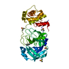
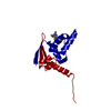
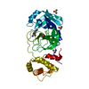
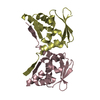
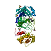
| Title | De novo design of protein homo-oligomers with modular hydrogen bond network-mediated specificity | ||||||
 Components Components | designed protein 3L6HC2_2 | ||||||
 Keywords Keywords | DE NOVO PROTEIN / rosetta / de novo design | ||||||
| Biological species | synthetic construct (others) | ||||||
| Method |  X-RAY DIFFRACTION / SYNCHROTRON / MOLECULAR REPLACEMENT / Resolution: 2.63 Å X-RAY DIFFRACTION / SYNCHROTRON / MOLECULAR REPLACEMENT / Resolution: 2.63 Å | ||||||
 Authors Authors | Sankaran, B. / Zwart, P.H. / Pereira, J.H. / Baker, D. / Boyken, S. / Chen, Z. / Groves, B. / Langan, R.A. / Oberdorfer, G. / Ford, A. ...Sankaran, B. / Zwart, P.H. / Pereira, J.H. / Baker, D. / Boyken, S. / Chen, Z. / Groves, B. / Langan, R.A. / Oberdorfer, G. / Ford, A. / Gilmore, J. / Xu, C. / DiMaio, F. / Seelig, G. | ||||||
 Citation Citation | Journal: Science / Year: 2016 Title: De novo design of protein homo-oligomers with modular hydrogen-bond network-mediated specificity. Authors: Boyken, S.E. / Chen, Z. / Groves, B. / Langan, R.A. / Oberdorfer, G. / Ford, A. / Gilmore, J.M. / Xu, C. / DiMaio, F. / Pereira, J.H. / Sankaran, B. / Seelig, G. / Zwart, P.H. / Baker, D. | ||||||
| History |
|
- Structure visualization
| Structure viewer | Molecule:  MolmilJmol/JSmol MolmilJmol/JSmol |
|---|
- Downloads & links
-Download
| PDBx/mmCIF format | 5j0l.cif.gz | 144.8 KB | Display | PDBx/mmCIF format |
|---|---|---|---|---|
| PDB format | pdb5j0l.ent.gz | 118.5 KB | Display | PDB format |
| PDBx/mmJSON format | 5j0l.json.gz | Tree view | PDBx/mmJSON format | |
| Others |  Other downloads Other downloads |
-Validation report
| Arichive directory | https://data.pdbj.org/pub/pdb/validation_reports/j0/5j0lftp://data.pdbj.org/pub/pdb/validation_reports/j0/5j0l | HTTPS FTP |
|---|
-Related structure data
| Related structure data |  5izsC  5j0hC  5j0iC  5j0jC  5j0kC  5j10C  5j2lC  5j73C  5jqzC C: citing same article ( |
|---|---|
| Similar structure data |

-Links
 PDBj
PDBj
- Assembly
| Deposited unit | 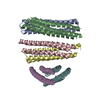
| ||||||||
|---|---|---|---|---|---|---|---|---|---|
| 1 | 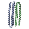
| ||||||||
| 2 | 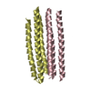
| ||||||||
| 3 | 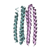
| ||||||||
| Unit cell |
|
-Components
| #1: Protein | Mass: 15969.917 Da / Num. of mol.: 6 Source method: isolated from a genetically manipulated source Source: (gene. exp.) synthetic construct (others) / Production host:  #2: Water | ChemComp-HOH / |  Mass: 18.015 Da / Num. of mol.: 131 / Source method: isolated from a natural source / Formula: H2O Mass: 18.015 Da / Num. of mol.: 131 / Source method: isolated from a natural source / Formula: H2O |
|---|
-Experimental details
-Experiment
| Experiment | Method: X-RAY DIFFRACTION / Number of used crystals: 1 |
|---|
- Sample preparation
| Crystal | Density Matthews: 2.69 Å3/Da / Density % sol: 54.3 % |
|---|---|
| Crystal grow | Temperature: 293 K / Method: vapor diffusion, sitting drop Details: 0.1 M Sodium Acetate trihydrate pH 4.5, 3.0 M Sodium chloride |
-Data collection
| Diffraction | Mean temperature: 100 K |
|---|---|
| Diffraction source | Source: SYNCHROTRON / Site: ALS  / Beamline: 8.2.2 / Wavelength: 1 Å / Beamline: 8.2.2 / Wavelength: 1 Å |
| Detector | Type: ADSC QUANTUM 315r / Detector: CCD / Date: Jan 28, 2015 |
| Radiation | Protocol: SINGLE WAVELENGTH / Monochromatic (M) / Laue (L): M / Scattering type: x-ray |
| Radiation wavelength | Wavelength: 1 Å / Relative weight: 1 |
| Reflection | Resolution: 2.63→66.1 Å / Num. obs: 27911 / % possible obs: 99.97 % / Redundancy: 12.5 % / Net I/σ(I): 17.9 |
- Processing
| Software |
| |||||||||||||||||||||||||||||||||||||||||||||||||||||||||||||||||||||||||||||||||||||||||||||||||||||||||
|---|---|---|---|---|---|---|---|---|---|---|---|---|---|---|---|---|---|---|---|---|---|---|---|---|---|---|---|---|---|---|---|---|---|---|---|---|---|---|---|---|---|---|---|---|---|---|---|---|---|---|---|---|---|---|---|---|---|---|---|---|---|---|---|---|---|---|---|---|---|---|---|---|---|---|---|---|---|---|---|---|---|---|---|---|---|---|---|---|---|---|---|---|---|---|---|---|---|---|---|---|---|---|---|---|---|---|
| Refinement | Method to determine structure: MOLECULAR REPLACEMENT / Resolution: 2.63→66.099 Å / SU ML: 0.78 / Cross valid method: THROUGHOUT / σ(F): 1.51 / Phase error: 30.27 / Stereochemistry target values: ML
| |||||||||||||||||||||||||||||||||||||||||||||||||||||||||||||||||||||||||||||||||||||||||||||||||||||||||
| Solvent computation | Shrinkage radii: 0.86 Å / VDW probe radii: 1.1 Å / Solvent model: FLAT BULK SOLVENT MODEL / Bsol: 41.217 Å2 / ksol: 0.334 e/Å3 | |||||||||||||||||||||||||||||||||||||||||||||||||||||||||||||||||||||||||||||||||||||||||||||||||||||||||
| Displacement parameters |
| |||||||||||||||||||||||||||||||||||||||||||||||||||||||||||||||||||||||||||||||||||||||||||||||||||||||||
| Refinement step | Cycle: LAST / Resolution: 2.63→66.099 Å
| |||||||||||||||||||||||||||||||||||||||||||||||||||||||||||||||||||||||||||||||||||||||||||||||||||||||||
| Refine LS restraints |
| |||||||||||||||||||||||||||||||||||||||||||||||||||||||||||||||||||||||||||||||||||||||||||||||||||||||||
| LS refinement shell |
|
