 Movie
Movie Controller

[English] 日本語
 Yorodumi
- PDB-1god: MONOMERIC LYS-49 PHOSPHOLIPASE A2 HOMOLOGUE ISOLATED FROM THE VEN... -
+ Open data

- Basic information
| Entry | Database: PDB / ID: 1god | ||||||
|---|---|---|---|---|---|---|---|
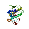
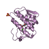
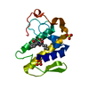
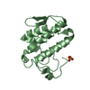
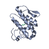
| Title | MONOMERIC LYS-49 PHOSPHOLIPASE A2 HOMOLOGUE ISOLATED FROM THE VENOM OF CERROPHIDION (BOTHROPS) GODMANI | ||||||
 Components Components | PROTEIN (PHOSPHOLIPASE A2) | ||||||
 Keywords Keywords | HYDROLASE / LYS49-PHOSPHOLIPASE A2 / SNAKE VENOM / BOTHROPS | ||||||
| Function / homology |  Function and homology information Function and homology informationA2-type glycerophospholipase activity / arachidonate secretion / lipid catabolic process / negative regulation of T cell proliferation / phospholipid metabolic process / phospholipid binding / toxin activity / calcium ion binding / extracellular region Similarity search - Function | ||||||
| Biological species |  Cerrophidion godmani (Godman's montane pit viper) Cerrophidion godmani (Godman's montane pit viper) | ||||||
| Method |  X-RAY DIFFRACTION / MOLECULAR REPLACEMENT / Resolution: 2.8 Å X-RAY DIFFRACTION / MOLECULAR REPLACEMENT / Resolution: 2.8 Å | ||||||
 Authors Authors | Arni, R.K. / Fontes, M.R.M. / Barberato, C. / Gutierrez, J.M. / Diaz-Oreiro, C. / Ward, R.J. | ||||||
 Citation Citation | Journal: Arch.Biochem.Biophys. / Year: 1999 Title: Crystal structure of myotoxin II, a monomeric Lys49-phospholipase A2 homologue isolated from the venom of Cerrophidion (Bothrops) godmani. Authors: Arni, R.K. / Fontes, M.R. / Barberato, C. / Gutierrez, J.M. / Diaz, C. / Ward, R.J. #1: Journal: Protein Pept.Lett. / Year: 1998Title: Crystallization and Preliminary X-Ray Diffraction Analysis of a Lys49-Pla2 Homologue from Cerrophidion Godmani Venom Authors: De Azevedo Jr., W.F. / Ward, R.J. / Gutierrez, J.M. / Diaz-Oreiro, C. / Arni, R.K. | ||||||
| History |
|
- Structure visualization
| Structure viewer | Molecule: MolmilJmol/JSmol |
|---|
- Downloads & links
-Download
| PDBx/mmCIF format | 1god.cif.gz | 37.2 KB | Display | PDBx/mmCIF format |
|---|---|---|---|---|
| PDB format | pdb1god.ent.gz | 25.3 KB | Display | PDB format |
| PDBx/mmJSON format | 1god.json.gz | Tree view | PDBx/mmJSON format | |
| Others |  Other downloads Other downloads |
-Validation report
| Arichive directory | https://data.pdbj.org/pub/pdb/validation_reports/go/1godftp://data.pdbj.org/pub/pdb/validation_reports/go/1god | HTTPS FTP |
|---|
-Related structure data
| Related structure data |  1clpS S: Starting model for refinement |
|---|---|
| Similar structure data |


-Links
 PDBj
PDBj
- Assembly
| Deposited unit | 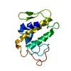
| ||||||||||
|---|---|---|---|---|---|---|---|---|---|---|---|
| 1 |
| ||||||||||
| Unit cell |
|
-Components
| #1: Protein | Mass: 13733.950 Da / Num. of mol.: 1 / Source method: isolated from a natural source Source: (natural) Cerrophidion godmani (Godman's montane pit viper)References: UniProt: P81165, phospholipase A2 |
|---|---|
| #2: Water | ChemComp-HOH /  Mass: 18.015 Da / Num. of mol.: 50 / Source method: isolated from a natural source / Formula: H2O Mass: 18.015 Da / Num. of mol.: 50 / Source method: isolated from a natural source / Formula: H2O |
| Has protein modification | Y |
-Experimental details
-Experiment
| Experiment | Method: X-RAY DIFFRACTION / Number of used crystals: 1 |
|---|
- Sample preparation
| Crystal | Density Matthews: 2.8 Å3/Da / Density % sol: 59 % | |||||||||||||||||||||||||||||||||||
|---|---|---|---|---|---|---|---|---|---|---|---|---|---|---|---|---|---|---|---|---|---|---|---|---|---|---|---|---|---|---|---|---|---|---|---|---|
| Crystal grow | Method: vapor diffusion, hanging drop / pH: 4.6 Details: 0.1 M SODIUM ACETATE (PH 4.6), 2.0 M (NH4)2SO4, 1MM NANO3, VAPOR DIFFUSION, HANGING DROP | |||||||||||||||||||||||||||||||||||
| Crystal | *PLUS | |||||||||||||||||||||||||||||||||||
| Crystal grow | *PLUS | |||||||||||||||||||||||||||||||||||
| Components of the solutions | *PLUS
|
-Data collection
| Diffraction | Mean temperature: 293 K |
|---|---|
| Diffraction source | Source: ROTATING ANODE / Type: RIGAKU RU200 / Wavelength: 1.5418 |
| Detector | Type: SIEMENS / Detector: AREA DETECTOR / Date: Jan 15, 1995 |
| Radiation | Monochromator: GRAPHITE / Protocol: SINGLE WAVELENGTH / Monochromatic (M) / Laue (L): M / Scattering type: x-ray |
| Radiation wavelength | Wavelength: 1.5418 Å / Relative weight: 1 |
| Reflection | Resolution: 2.8→8 Å / Num. obs: 22540 / % possible obs: 99.5 % / Observed criterion σ(I): 2 / Redundancy: 3 % / Rsym value: 0.09 |
| Reflection | *PLUS Num. obs: 4482 / Num. measured all: 22540 / Rmerge(I) obs: 0.09 |
- Processing
| Software |
| ||||||||||||||||||||||||||||||||||||||||||||||||||||||||||||
|---|---|---|---|---|---|---|---|---|---|---|---|---|---|---|---|---|---|---|---|---|---|---|---|---|---|---|---|---|---|---|---|---|---|---|---|---|---|---|---|---|---|---|---|---|---|---|---|---|---|---|---|---|---|---|---|---|---|---|---|---|---|
| Refinement | Method to determine structure: MOLECULAR REPLACEMENT Starting model: 1CLP Resolution: 2.8→8 Å / Rfactor Rfree error: 0.016 / Data cutoff high absF: 10000000 / Data cutoff low absF: 0.001 / Isotropic thermal model: OVERALL / Cross valid method: THROUGHOUT / σ(F): 2
| ||||||||||||||||||||||||||||||||||||||||||||||||||||||||||||
| Displacement parameters | Biso mean: 35.5 Å2 | ||||||||||||||||||||||||||||||||||||||||||||||||||||||||||||
| Refine analyze |
| ||||||||||||||||||||||||||||||||||||||||||||||||||||||||||||
| Refinement step | Cycle: LAST / Resolution: 2.8→8 Å
| ||||||||||||||||||||||||||||||||||||||||||||||||||||||||||||
| Refine LS restraints |
| ||||||||||||||||||||||||||||||||||||||||||||||||||||||||||||
| LS refinement shell | Resolution: 2.8→2.97 Å / Rfactor Rfree error: 0.068 / Total num. of bins used: 6
| ||||||||||||||||||||||||||||||||||||||||||||||||||||||||||||
| Xplor file |
| ||||||||||||||||||||||||||||||||||||||||||||||||||||||||||||
| Software | *PLUS Name: X-PLOR / Version: 3.851 / Classification: refinement | ||||||||||||||||||||||||||||||||||||||||||||||||||||||||||||
| Refinement | *PLUS Highest resolution: 2.8 Å / Lowest resolution: 8 Å / σ(F): 2 / % reflection Rfree: 10.5 % / Rfactor obs: 0.188 | ||||||||||||||||||||||||||||||||||||||||||||||||||||||||||||
| Solvent computation | *PLUS | ||||||||||||||||||||||||||||||||||||||||||||||||||||||||||||
| Displacement parameters | *PLUS Biso mean: 35.5 Å2 | ||||||||||||||||||||||||||||||||||||||||||||||||||||||||||||
| Refine LS restraints | *PLUS
| ||||||||||||||||||||||||||||||||||||||||||||||||||||||||||||
| LS refinement shell | *PLUS Highest resolution: 2.8 Å / Rfactor Rfree: 0.36 / % reflection Rfree: 11.4 % / Rfactor Rwork: 0.23 |
