- PDB-3ux7: Crystal structure of a dimeric myotoxic component of the Vipera a... -
+
Open data
ID or keywords:
Loading...
-
Basic information
Entry
Database: PDB / ID: 3ux7
Title
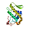
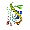
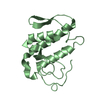
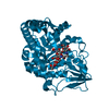
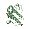
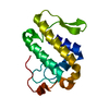
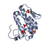
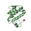
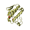
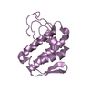
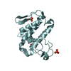
Crystal structure of a dimeric myotoxic component of the Vipera ammodytes meridionalis venom reveals determinants of myotoxicity and membrane damaging activity
Components
Ammodytin L(1) isoform
Keywords
TOXIN / Snake venom / Ser49 myotoxin
Function / homology
Function and homology information
: / arachidonate secretion / lipid catabolic process / negative regulation of T cell proliferation / phospholipid metabolic process / phospholipid binding / toxin activity / calcium ion binding / extracellular region Similarity search - Function
Phospholipase A2, aspartic acid active site / Phospholipase A2 aspartic acid active site. / Phospholipase A2, histidine active site / Phospholipase A2 histidine active site. / Phospholipase A2 / Phospholipase A2 domain / Phospholipase A2 / Phospholipase A2 / Phospholipase A2 domain / Phospholipase A2 ...Phospholipase A2, aspartic acid active site / Phospholipase A2 aspartic acid active site. / Phospholipase A2, histidine active site / Phospholipase A2 histidine active site. / Phospholipase A2 / Phospholipase A2 domain / Phospholipase A2 / Phospholipase A2 / Phospholipase A2 domain / Phospholipase A2 / Phospholipase A2 domain superfamily / Up-down Bundle / Mainly Alpha Similarity search - Domain/homology
Mass: 18.015 Da / Num. of mol.: 90 / Source method: isolated from a natural source / Formula: H2O
Has protein modification
Y
-
Experimental details
-
Experiment
Experiment
Method: X-RAY DIFFRACTION / Number of used crystals: 1
-
Sample preparation
Crystal
Density Matthews: 2.16 Å3/Da / Density % sol: 42.93 %
Crystal grow
Method: vapor diffusion, hanging drop Details: Single crystals suitable for X-ray analysis were obtained after 5 days by the hanging-drop vapour-diffusion technique, using a protein solution with concentration of 10mg/ml in water. The ...Details: Single crystals suitable for X-ray analysis were obtained after 5 days by the hanging-drop vapour-diffusion technique, using a protein solution with concentration of 10mg/ml in water. The reservoir solution contained 25.5% PEG-4000, 0.17 M ammonium sulphate and 15% glycerol., VAPOR DIFFUSION, HANGING DROP
Protocol: SINGLE WAVELENGTH / Monochromatic (M) / Laue (L): M / Scattering type: x-ray
Radiation wavelength
Wavelength: 0.8123 Å / Relative weight: 1
Reflection
Resolution: 2.95→40 Å / Num. obs: 21213
-
Processing
Software
Name
Version
Classification
DNA
datacollection
PHASER
phasing
REFMAC
5.6.0117
refinement
DENZO
datareduction
SCALEPACK
datascaling
Refinement
Method to determine structure: MOLECULAR REPLACEMENT / Resolution: 2.97→40 Å / Cor.coef. Fo:Fc: 0.94 / Cor.coef. Fo:Fc free: 0.914 / SU B: 15.611 / SU ML: 0.301 / Cross valid method: THROUGHOUT / ESU R Free: 0.52 / Stereochemistry target values: MAXIMUM LIKELIHOOD / Details: HYDROGENS HAVE BEEN USED IF PRESENT IN THE INPUT
Rfactor
Num. reflection
% reflection
Selection details
Rfree
0.26079
972
5.1 %
RANDOM
Rwork
0.22439
-
-
-
obs
0.22626
17915
97.05 %
-
Solvent computation
Ion probe radii: 0.8 Å / Shrinkage radii: 0.8 Å / VDW probe radii: 1.2 Å / Solvent model: MASK
Displacement parameters
Biso mean: 27.764 Å2
Baniso -1
Baniso -2
Baniso -3
1-
3.77 Å2
-0 Å2
-0.61 Å2
2-
-
-1.71 Å2
0 Å2
3-
-
-
-2.07 Å2
Refinement step
Cycle: LAST / Resolution: 2.97→40 Å
Protein
Nucleic acid
Ligand
Solvent
Total
Num. atoms
7463
0
95
90
7648
Refine LS restraints
Refine-ID
Type
Dev ideal
Dev ideal target
Number
X-RAY DIFFRACTION
r_bond_refined_d
0.008
0.02
7728
X-RAY DIFFRACTION
r_bond_other_d
X-RAY DIFFRACTION
r_angle_refined_deg
1.279
1.973
10398
X-RAY DIFFRACTION
r_angle_other_deg
X-RAY DIFFRACTION
r_dihedral_angle_1_deg
5.876
5
968
X-RAY DIFFRACTION
r_dihedral_angle_2_deg
37.622
24.153
301
X-RAY DIFFRACTION
r_dihedral_angle_3_deg
18.532
15
1360
X-RAY DIFFRACTION
r_dihedral_angle_4_deg
20.067
15
24
X-RAY DIFFRACTION
r_chiral_restr
0.085
0.2
1071
X-RAY DIFFRACTION
r_gen_planes_refined
0.004
0.021
5684
X-RAY DIFFRACTION
r_gen_planes_other
X-RAY DIFFRACTION
r_nbd_refined
X-RAY DIFFRACTION
r_nbd_other
X-RAY DIFFRACTION
r_nbtor_refined
X-RAY DIFFRACTION
r_nbtor_other
X-RAY DIFFRACTION
r_xyhbond_nbd_refined
X-RAY DIFFRACTION
r_xyhbond_nbd_other
X-RAY DIFFRACTION
r_metal_ion_refined
X-RAY DIFFRACTION
r_metal_ion_other
X-RAY DIFFRACTION
r_symmetry_vdw_refined
X-RAY DIFFRACTION
r_symmetry_vdw_other
X-RAY DIFFRACTION
r_symmetry_hbond_refined
X-RAY DIFFRACTION
r_symmetry_hbond_other
X-RAY DIFFRACTION
r_symmetry_metal_ion_refined
X-RAY DIFFRACTION
r_symmetry_metal_ion_other
X-RAY DIFFRACTION
r_mcbond_it
X-RAY DIFFRACTION
r_mcbond_other
X-RAY DIFFRACTION
r_mcangle_it
X-RAY DIFFRACTION
r_scbond_it
X-RAY DIFFRACTION
r_scangle_it
X-RAY DIFFRACTION
r_rigid_bond_restr
X-RAY DIFFRACTION
r_sphericity_free
X-RAY DIFFRACTION
r_sphericity_bonded
LS refinement shell
Resolution: 2.97→3.047 Å / Total num. of bins used: 20
Rfactor
Num. reflection
% reflection
Rfree
0.333
75
-
Rwork
0.269
1197
-
obs
-
-
96.07 %
+
About Yorodumi
-
News
-
Feb 9, 2022. New format data for meta-information of EMDB entries
New format data for meta-information of EMDB entries
Version 3 of the EMDB header file is now the official format.
The previous official version 1.9 will be removed from the archive.
In the structure databanks used in Yorodumi, some data are registered as the other names, "COVID-19 virus" and "2019-nCoV". Here are the details of the virus and the list of structure data.
Jan 31, 2019. EMDB accession codes are about to change! (news from PDBe EMDB page)
EMDB accession codes are about to change! (news from PDBe EMDB page)
The allocation of 4 digits for EMDB accession codes will soon come to an end. Whilst these codes will remain in use, new EMDB accession codes will include an additional digit and will expand incrementally as the available range of codes is exhausted. The current 4-digit format prefixed with “EMD-” (i.e. EMD-XXXX) will advance to a 5-digit format (i.e. EMD-XXXXX), and so on. It is currently estimated that the 4-digit codes will be depleted around Spring 2019, at which point the 5-digit format will come into force.
The EM Navigator/Yorodumi systems omit the EMD- prefix.
Related info.:Q: What is EMD? / ID/Accession-code notation in Yorodumi/EM Navigator
Yorodumi is a browser for structure data from EMDB, PDB, SASBDB, etc.
This page is also the successor to EM Navigator detail page, and also detail information page/front-end page for Omokage search.
The word "yorodu" (or yorozu) is an old Japanese word meaning "ten thousand". "mi" (miru) is to see.
Related info.:EMDB / PDB / SASBDB / Comparison of 3 databanks / Yorodumi Search / Aug 31, 2016. New EM Navigator & Yorodumi / Yorodumi Papers / Jmol/JSmol / Function and homology information / Changes in new EM Navigator and Yorodumi
 Movie
Movie Controller

 Yorodumi
 Open data
 Basic information
 Components
 Keywords
 Function and homology information
 Vipera ammodytes meridionalis (snake)
 X-RAY DIFFRACTION /
  Authors
 Citation
 Structure visualization
 Downloads & links
 Other downloads
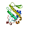


 PDBj
PDBj
 Assembly


 Mass: 96.063 Da / Num. of mol.: 19 / Source method: obtained synthetically / Formula: SO4
 Mass: 18.015 Da / Num. of mol.: 90 / Source method: isolated from a natural source / Formula: H2O
 Sample preparation
 / Beamline: X13 / Wavelength: 0.8123 Å
 Processing