 Movie
Movie Controller

[English] 日本語
 Yorodumi
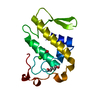
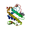
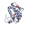
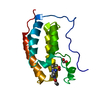
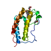
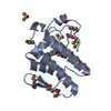
- PDB-1m8s: Crystal Structures of Cadmium-binding Acidic Phospholipase A2 fro... -
+ Open data

- Basic information
| Entry | Database: PDB / ID: 1m8s | ||||||
|---|---|---|---|---|---|---|---|
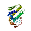
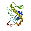
| Title | Crystal Structures of Cadmium-binding Acidic Phospholipase A2 from the Venom of Agkistrodon halys pallas at 1.9 Resolution (crystal grown at pH 5.9) | ||||||
 Components Components | phospholipase a2 | ||||||
 Keywords Keywords | HYDROLASE / phopholipase a2-metal cation complex / three alpha / two beta | ||||||
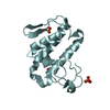
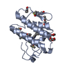
| Function / homology |  Function and homology information Function and homology informationphospholipase A2 / : / arachidonate secretion / lipid catabolic process / negative regulation of T cell proliferation / phospholipid metabolic process / phospholipid binding / toxin activity / calcium ion binding / extracellular region Similarity search - Function | ||||||
| Biological species |  Gloydius halys (Halys viper) Gloydius halys (Halys viper) | ||||||
| Method |  X-RAY DIFFRACTION / isomorphous difference Fourier / Resolution: 1.9 Å X-RAY DIFFRACTION / isomorphous difference Fourier / Resolution: 1.9 Å | ||||||
 Authors Authors | Xu, S. / Gu, L. / Zhou, Y. / Lin, Z. | ||||||
 Citation Citation | Journal: Biochem.Biophys.Res.Commun. / Year: 2003 Title: Structures of cadmium-binding acidic phospholipase A(2) from the venom of Agkistrodon halys Pallas at 1.9A resolutio Authors: Xu, S. / Gu, L. / Jiang, T. / Zhou, Y. / Lin, Z. | ||||||
| History |
|
- Structure visualization
| Structure viewer | Molecule: MolmilJmol/JSmol |
|---|
- Downloads & links
-Download
| PDBx/mmCIF format | 1m8s.cif.gz | 40.8 KB | Display | PDBx/mmCIF format |
|---|---|---|---|---|
| PDB format | pdb1m8s.ent.gz | 27.3 KB | Display | PDB format |
| PDBx/mmJSON format | 1m8s.json.gz | Tree view | PDBx/mmJSON format | |
| Others |  Other downloads Other downloads |
-Validation report
| Arichive directory | https://data.pdbj.org/pub/pdb/validation_reports/m8/1m8sftp://data.pdbj.org/pub/pdb/validation_reports/m8/1m8s | HTTPS FTP |
|---|
-Related structure data
| Related structure data |  1m8rC 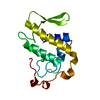 1psjS S: Starting model for refinement C: citing same article ( |
|---|---|
| Similar structure data |

-Links
 PDBj
PDBj


- Assembly
| Deposited unit | 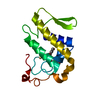
| ||||||||
|---|---|---|---|---|---|---|---|---|---|
| 1 |
| ||||||||
| Unit cell |
|
-Components
| #1: Protein | Mass: 13956.688 Da / Num. of mol.: 1 / Source method: isolated from a natural source / Source: (natural) Gloydius halys (Halys viper) / Secretion: venomReferences: UniProt: P14418, UniProt: O42191*PLUS, phospholipase A2 | ||||
|---|---|---|---|---|---|
| #2: Chemical | ChemComp-CD /   Mass: 112.411 Da / Num. of mol.: 1 / Source method: obtained synthetically / Formula: Cd Mass: 112.411 Da / Num. of mol.: 1 / Source method: obtained synthetically / Formula: Cd | ||||
| #3: Chemical | 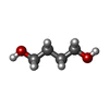  Mass: 90.121 Da / Num. of mol.: 2 / Source method: obtained synthetically / Formula: C4H10O2 Mass: 90.121 Da / Num. of mol.: 2 / Source method: obtained synthetically / Formula: C4H10O2#4: Water | ChemComp-HOH / |  Mass: 18.015 Da / Num. of mol.: 110 / Source method: isolated from a natural source / Formula: H2O Mass: 18.015 Da / Num. of mol.: 110 / Source method: isolated from a natural source / Formula: H2OHas protein modification | Y | |
-Experimental details
-Experiment
| Experiment | Method: X-RAY DIFFRACTION / Number of used crystals: 1 |
|---|
- Sample preparation
| Crystal | Density Matthews: 2.34 Å3/Da / Density % sol: 47.52 % | ||||||||||||||||||||||||||||||||||||||||||
|---|---|---|---|---|---|---|---|---|---|---|---|---|---|---|---|---|---|---|---|---|---|---|---|---|---|---|---|---|---|---|---|---|---|---|---|---|---|---|---|---|---|---|---|
| Crystal grow | Temperature: 293 K / Method: vapor diffusion, hanging drop / pH: 5.9 Details: 10%(v/v) 1,4-butyldiol, 0.01M CdCl2, 0.1M Na(CH3)2AsO, pH 5.9, VAPOR DIFFUSION, HANGING DROP, temperature 293K | ||||||||||||||||||||||||||||||||||||||||||
| Crystal grow | *PLUS Temperature: 291 K | ||||||||||||||||||||||||||||||||||||||||||
| Components of the solutions | *PLUS
|
-Data collection
| Diffraction | Mean temperature: 293 K |
|---|---|
| Diffraction source | Source: ROTATING ANODE / Type: RIGAKU / Wavelength: 1.5418 Å |
| Detector | Type: MARRESEARCH / Detector: AREA DETECTOR / Date: Jan 11, 2001 |
| Radiation | Monochromator: graphite / Protocol: SINGLE WAVELENGTH / Monochromatic (M) / Laue (L): M / Scattering type: x-ray |
| Radiation wavelength | Wavelength: 1.5418 Å / Relative weight: 1 |
| Reflection | Resolution: 1.9→25 Å / Num. all: 10431 / Num. obs: 10431 / % possible obs: 100 % / Observed criterion σ(I): 0 / Redundancy: 9.8 % / Biso Wilson estimate: 17.8 Å2 / Rmerge(I) obs: 0.072 / Net I/σ(I): 33.8 |
| Reflection shell | Resolution: 1.9→1.97 Å / Redundancy: 9.4 % / Rmerge(I) obs: 0.26 / Mean I/σ(I) obs: 9.7 / Num. unique all: 1024 / % possible all: 100 |
| Reflection | *PLUS Lowest resolution: 25 Å / % possible obs: 100 % / Num. measured all: 187637 |
| Reflection shell | *PLUS % possible obs: 100 % / Rmerge(I) obs: 0.26 |
- Processing
| Software |
| ||||||||||||||||||||||||||||||||||||
|---|---|---|---|---|---|---|---|---|---|---|---|---|---|---|---|---|---|---|---|---|---|---|---|---|---|---|---|---|---|---|---|---|---|---|---|---|---|
| Refinement | Method to determine structure: isomorphous difference Fourier Starting model: PDB ENTRY 1PSJ Resolution: 1.9→25 Å / Rfactor Rfree error: 0.006 / Isotropic thermal model: RESTRAINED / Cross valid method: THROUGHOUT / σ(F): 0
| ||||||||||||||||||||||||||||||||||||
| Solvent computation | Solvent model: FLAT MODEL / Bsol: 126.972 Å2 / ksol: 0.44155 e/Å3 | ||||||||||||||||||||||||||||||||||||
| Displacement parameters | Biso mean: 22.3 Å2
| ||||||||||||||||||||||||||||||||||||
| Refine analyze | Luzzati coordinate error free: 0.2 Å / Luzzati sigma a free: 0.08 Å | ||||||||||||||||||||||||||||||||||||
| Refinement step | Cycle: LAST / Resolution: 1.9→25 Å
| ||||||||||||||||||||||||||||||||||||
| Refine LS restraints |
| ||||||||||||||||||||||||||||||||||||
| LS refinement shell | Resolution: 1.9→2.02 Å / Rfactor Rfree error: 0.02 / Total num. of bins used: 6
| ||||||||||||||||||||||||||||||||||||
| Xplor file |
| ||||||||||||||||||||||||||||||||||||
| Refinement | *PLUS Highest resolution: 1.9 Å / Lowest resolution: 25 Å / Rfactor Rfree: 0.208 / Rfactor Rwork: 0.198 | ||||||||||||||||||||||||||||||||||||
| Solvent computation | *PLUS | ||||||||||||||||||||||||||||||||||||
| Displacement parameters | *PLUS | ||||||||||||||||||||||||||||||||||||
| Refine LS restraints | *PLUS
|
