 Movie
Movie Controller

[English] 日本語
 Yorodumi
- PDB-1xx9: Crystal Structure of the FXIa Catalytic Domain in Complex with Ec... -
+ Open data

- Basic information
| Entry | Database: PDB / ID: 1xx9 | ||||||
|---|---|---|---|---|---|---|---|
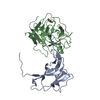
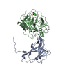
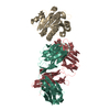
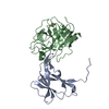
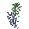
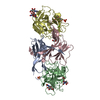
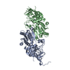
| Title | Crystal Structure of the FXIa Catalytic Domain in Complex with EcotinM84R | ||||||
 Components Components |
| ||||||
 Keywords Keywords | BLOOD CLOTTING/HYDROLASE INHIBITOR / FXIa / Catalytic domain / Serine protein / Ecotin / BLOOD CLOTTING-HYDROLASE INHIBITOR COMPLEX | ||||||
| Function / homology |  Function and homology information Function and homology informationcoagulation factor XIa / serine-type aminopeptidase activity / Defective F9 activation / positive regulation of fibrinolysis / plasminogen activation / : / serine-type peptidase activity / serine-type endopeptidase inhibitor activity / defense response / blood coagulation ...coagulation factor XIa / serine-type aminopeptidase activity / Defective F9 activation / positive regulation of fibrinolysis / plasminogen activation / : / serine-type peptidase activity / serine-type endopeptidase inhibitor activity / defense response / blood coagulation / heparin binding / outer membrane-bounded periplasmic space / serine-type endopeptidase activity / protein homodimerization activity / : / extracellular exosome / extracellular region / membrane / identical protein binding / plasma membrane Similarity search - Function | ||||||
| Biological species |  Homo sapiens (human) Homo sapiens (human) | ||||||
| Method |  X-RAY DIFFRACTION / SYNCHROTRON / MOLECULAR REPLACEMENT / Resolution: 2.2 Å X-RAY DIFFRACTION / SYNCHROTRON / MOLECULAR REPLACEMENT / Resolution: 2.2 Å | ||||||
 Authors Authors | Jin, L. / Pandey, P. / Babine, R.E. / Gorga, J.C. / Seidl, K.J. / Gelfand, E. / Weaver, D.T. / Abdel-Meguid, S.S. / Strickler, J.E. | ||||||
 Citation Citation | Journal: J.Biol.Chem. / Year: 2005 Title: Crystal Structures of the FXIa Catalytic Domain in Complex with Ecotin Mutants Reveal Substrate-like Interactions Authors: Jin, L. / Pandey, P. / Babine, R.E. / Gorga, J.C. / Seidl, K.J. / Gelfand, E. / Weaver, D.T. / Abdel-Meguid, S.S. / Strickler, J.E. | ||||||
| History |
|
- Structure visualization
| Structure viewer | Molecule: MolmilJmol/JSmol |
|---|
- Downloads & links
-Download
| PDBx/mmCIF format | 1xx9.cif.gz | 165.4 KB | Display | PDBx/mmCIF format |
|---|---|---|---|---|
| PDB format | pdb1xx9.ent.gz | 130.2 KB | Display | PDB format |
| PDBx/mmJSON format | 1xx9.json.gz | Tree view | PDBx/mmJSON format | |
| Others |  Other downloads Other downloads |
-Validation report
| Arichive directory | https://data.pdbj.org/pub/pdb/validation_reports/xx/1xx9ftp://data.pdbj.org/pub/pdb/validation_reports/xx/1xx9 | HTTPS FTP |
|---|
-Related structure data
| Related structure data |  1xxdC  1xxfC  1fi8S C: citing same article ( S: Starting model for refinement |
|---|---|
| Similar structure data |


-Links
 PDBj
PDBj


- Assembly
| Deposited unit | 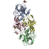
| ||||||||||
|---|---|---|---|---|---|---|---|---|---|---|---|
| 1 |
| ||||||||||
| Unit cell |
|
-Components
| #1: Protein | Mass: 26872.562 Da / Num. of mol.: 2 / Fragment: Catalytic Domain Source method: isolated from a genetically manipulated source Source: (gene. exp.) Homo sapiens (human) / Gene: F11 / Production host:  Pichia pastoris (fungus) / References: UniProt: P03951, coagulation factor XIa Pichia pastoris (fungus) / References: UniProt: P03951, coagulation factor XIa#2: Protein | Mass: 16146.505 Da / Num. of mol.: 2 / Mutation: M84R Source method: isolated from a genetically manipulated source Source: (gene. exp.) #3: Sugar | ChemComp-NAG / |   Type: D-saccharide, beta linking / Mass: 221.208 Da / Num. of mol.: 1 Type: D-saccharide, beta linking / Mass: 221.208 Da / Num. of mol.: 1Source method: isolated from a genetically manipulated source Formula: C8H15NO6 #4: Water | ChemComp-HOH / |  Mass: 18.015 Da / Num. of mol.: 260 / Source method: isolated from a natural source / Formula: H2O Mass: 18.015 Da / Num. of mol.: 260 / Source method: isolated from a natural source / Formula: H2OHas protein modification | Y | |
|---|
-Experimental details
-Experiment
| Experiment | Method: X-RAY DIFFRACTION / Number of used crystals: 1 |
|---|
- Sample preparation
| Crystal | Density Matthews: 2.1 Å3/Da / Density % sol: 41 % |
|---|---|
| Crystal grow | Temperature: 293 K / Method: vapor diffusion, hanging drop / pH: 6.2 Details: PEG 1000, NaCl, Na/K phosphate, pH 6.2, VAPOR DIFFUSION, HANGING DROP, temperature 293K |
-Data collection
| Diffraction | Mean temperature: 113 K |
|---|---|
| Diffraction source | Source: SYNCHROTRON / Site: NSLS  / Beamline: X12C / Wavelength: 1.1 Å / Beamline: X12C / Wavelength: 1.1 Å |
| Detector | Type: CUSTOM-MADE / Detector: CCD / Date: Aug 9, 2001 / Details: monochromator |
| Radiation | Monochromator: channel-cut crystal / Protocol: SINGLE WAVELENGTH / Monochromatic (M) / Laue (L): M / Scattering type: x-ray |
| Radiation wavelength | Wavelength: 1.1 Å / Relative weight: 1 |
| Reflection | Resolution: 2.2→50 Å / Num. all: 40129 / Num. obs: 36197 / % possible obs: 90.2 % / Observed criterion σ(I): 2 / Redundancy: 4.2 % / Biso Wilson estimate: 9.4 Å2 / Rsym value: 0.073 / Net I/σ(I): 12.3 |
| Reflection shell | Resolution: 2.2→2.28 Å / Rmerge(I) obs: 0.386 / Mean I/σ(I) obs: 2 / Num. unique all: 2715 / % possible all: 69.1 |
- Processing
| Software |
| ||||||||||||||||||||||||||||||||||||
|---|---|---|---|---|---|---|---|---|---|---|---|---|---|---|---|---|---|---|---|---|---|---|---|---|---|---|---|---|---|---|---|---|---|---|---|---|---|
| Refinement | Method to determine structure: MOLECULAR REPLACEMENT Starting model: PDB Entry 1FI8 Resolution: 2.2→24.63 Å / Rfactor Rfree error: 0.005 / Data cutoff high absF: 263306.1 / Data cutoff low absF: 0 / Isotropic thermal model: RESTRAINED / Cross valid method: THROUGHOUT / σ(F): 0 / Stereochemistry target values: Engh & Huber
| ||||||||||||||||||||||||||||||||||||
| Solvent computation | Solvent model: FLAT MODEL / Bsol: 26.9924 Å2 / ksol: 0.333821 e/Å3 | ||||||||||||||||||||||||||||||||||||
| Displacement parameters | Biso mean: 39.5 Å2
| ||||||||||||||||||||||||||||||||||||
| Refine analyze |
| ||||||||||||||||||||||||||||||||||||
| Refinement step | Cycle: LAST / Resolution: 2.2→24.63 Å
| ||||||||||||||||||||||||||||||||||||
| Refine LS restraints |
| ||||||||||||||||||||||||||||||||||||
| LS refinement shell | Resolution: 2.2→2.28 Å / Rfactor Rfree error: 0.027 / Total num. of bins used: 10
| ||||||||||||||||||||||||||||||||||||
| Xplor file |
|
