 Movie
Movie Controller


+ Open data

- Basic information
| Entry | Database: PDB / ID: 1fi8 | ||||||
|---|---|---|---|---|---|---|---|
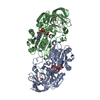
| Title | RAT GRANZYME B [N66Q] COMPLEXED TO ECOTIN [81-84 IEPD] | ||||||
 Components Components |
| ||||||
 Keywords Keywords | HYDROLASE/HYDROLASE INHIBITOR / complex (serine protease-inhibitor) / protease substrate interactions / beta strand structure / chymotrypsin fold / granzyme B / ecotin / HYDROLASE-HYDROLASE INHIBITOR COMPLEX | ||||||
| Function / homology |  Function and homology information Function and homology informationActivation, myristolyation of BID and translocation to mitochondria / granzyme B / Pyroptosis / positive regulation of immune response to tumor cell / granzyme-mediated programmed cell death signaling pathway / cytolytic granule / pyroptotic cell death / positive regulation of necroptotic process / plasma membrane repair / ceramide biosynthetic process ...Activation, myristolyation of BID and translocation to mitochondria / granzyme B / Pyroptosis / positive regulation of immune response to tumor cell / granzyme-mediated programmed cell death signaling pathway / cytolytic granule / pyroptotic cell death / positive regulation of necroptotic process / plasma membrane repair / ceramide biosynthetic process / natural killer cell mediated cytotoxicity / protein secretion / pyroptotic inflammatory response / serine-type peptidase activity / : / T cell mediated cytotoxicity / serine-type endopeptidase inhibitor activity / defense response / protein maturation / outer membrane-bounded periplasmic space / neuron apoptotic process / killing of cells of another organism / early endosome / defense response to bacterium / negative regulation of translation / serine-type endopeptidase activity / protein homodimerization activity / : / membrane / cytoplasm Similarity search - Function | ||||||
| Biological species |   | ||||||
| Method |  X-RAY DIFFRACTION / SYNCHROTRON / Resolution: 2.2 Å X-RAY DIFFRACTION / SYNCHROTRON / Resolution: 2.2 Å | ||||||
 Authors Authors | Waugh, S.M. / Harris, J.L. / Fletterick, R.J. / Craik, C.S. | ||||||
 Citation Citation | Journal: Nat.Struct.Biol. / Year: 2000 Title: The structure of the pro-apoptotic protease granzyme B reveals the molecular determinants of its specificity Authors: Waugh, S.M. / Harris, J.L. / Fletterick, R. / Craik, C.S. | ||||||
| History |
|
- Structure visualization
| Structure viewer | Molecule: MolmilJmol/JSmol |
|---|
- Downloads & links
-Download
| PDBx/mmCIF format | 1fi8.cif.gz | 153.3 KB | Display | PDBx/mmCIF format |
|---|---|---|---|---|
| PDB format | pdb1fi8.ent.gz | 118.9 KB | Display | PDB format |
| PDBx/mmJSON format | 1fi8.json.gz | Tree view | PDBx/mmJSON format | |
| Others |  Other downloads Other downloads |
-Validation report
| Arichive directory | https://data.pdbj.org/pub/pdb/validation_reports/fi/1fi8ftp://data.pdbj.org/pub/pdb/validation_reports/fi/1fi8 | HTTPS FTP |
|---|
-Related structure data
| Similar structure data |
|---|


-Links
 PDBj
PDBj- Assembly
| Deposited unit | 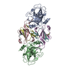
| ||||||||
|---|---|---|---|---|---|---|---|---|---|
| 1 |
| ||||||||
| Unit cell |
| ||||||||
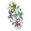
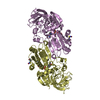
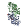
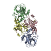
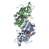
| Details | the biological assembly is the tetramer in the asymmetric unit. |
-Components
| #1: Protein | Mass: 25225.396 Da / Num. of mol.: 2 / Mutation: N66Q Source method: isolated from a genetically manipulated source Source: (gene. exp.)  Pichia pastoris (fungus) Pichia pastoris (fungus)References: UniProt: P18291, Hydrolases; Acting on peptide bonds (peptidases); Serine endopeptidases #2: Protein | Mass: 9528.733 Da / Num. of mol.: 2 / Fragment: RESIDUES 28 - 111 / Mutation: V81I, S82E, T83P, M84D Source method: isolated from a genetically manipulated source Source: (gene. exp.) #3: Protein | Mass: 6645.746 Da / Num. of mol.: 2 / Fragment: RESIDUES 112 - 169 Source method: isolated from a genetically manipulated source Source: (gene. exp.) #4: Water | ChemComp-HOH / |  Mass: 18.015 Da / Num. of mol.: 370 / Source method: isolated from a natural source / Formula: H2O Mass: 18.015 Da / Num. of mol.: 370 / Source method: isolated from a natural source / Formula: H2OHas protein modification | Y | |
|---|
-Experimental details
-Experiment
| Experiment | Method: X-RAY DIFFRACTION / Number of used crystals: 2 |
|---|
- Sample preparation
| Crystal | Density Matthews: 2.63 Å3/Da / Density % sol: 49.15 % | ||||||||||||||||||||||||||||||||||||||||||
|---|---|---|---|---|---|---|---|---|---|---|---|---|---|---|---|---|---|---|---|---|---|---|---|---|---|---|---|---|---|---|---|---|---|---|---|---|---|---|---|---|---|---|---|
| Crystal grow | Temperature: 298 K / Method: vapor diffusion, hanging drop / pH: 4.8 Details: sodium acetate, PEGmme 2000, ammonium acetate,, pH 4.8, VAPOR DIFFUSION, HANGING DROP, temperature 298K | ||||||||||||||||||||||||||||||||||||||||||
| Crystal grow | *PLUS Temperature: 18 ℃ / pH: 5.8 | ||||||||||||||||||||||||||||||||||||||||||
| Components of the solutions | *PLUS
|
-Data collection
| Diffraction |
| |||||||||||||||
|---|---|---|---|---|---|---|---|---|---|---|---|---|---|---|---|---|
| Diffraction source |
| |||||||||||||||
| Detector |
| |||||||||||||||
| Radiation | Protocol: SINGLE WAVELENGTH / Monochromatic (M) / Laue (L): M / Scattering type: x-ray | |||||||||||||||
| Radiation wavelength | Wavelength: 1.08 Å / Relative weight: 1 | |||||||||||||||
| Reflection | Resolution: 2.2→35.8 Å / Num. all: 98218 / Num. obs: 98218 / % possible obs: 90.3 % / Observed criterion σ(I): 1 / Redundancy: 2.7 % / Biso Wilson estimate: 35.1 Å2 / Rmerge(I) obs: 0.093 / Net I/σ(I): 6.8 | |||||||||||||||
| Reflection shell | Resolution: 2.21→2.28 Å / Redundancy: 2.7 % / Rmerge(I) obs: 0.58 / Num. unique all: 48837 / % possible all: 90.7 | |||||||||||||||
| Reflection | *PLUS Num. obs: 48837 / Num. measured all: 121975 | |||||||||||||||
| Reflection shell | *PLUS % possible obs: 90 % |

- Processing
| Software |
| |||||||||||||||||||||||||
|---|---|---|---|---|---|---|---|---|---|---|---|---|---|---|---|---|---|---|---|---|---|---|---|---|---|---|
| Refinement | Resolution: 2.2→35.7 Å / σ(F): 0 / σ(I): 0 / Stereochemistry target values: Engh and Huber Details: Crystal 1 data was used for initial molecular replacement and refinement. Crystal 2 data was given the same Rfree set and used for final refinement. Molecular replacement used 1AZZ and 1AUG.
| |||||||||||||||||||||||||
| Displacement parameters | Biso mean: 43.09 Å2 | |||||||||||||||||||||||||
| Refine analyze |
| |||||||||||||||||||||||||
| Refinement step | Cycle: LAST / Resolution: 2.2→35.7 Å
| |||||||||||||||||||||||||
| Refine LS restraints |
| |||||||||||||||||||||||||
| Software | *PLUS Name: CNS / Version: 0.9 / Classification: refinement | |||||||||||||||||||||||||
| Refinement | *PLUS Highest resolution: 2.2 Å / Lowest resolution: 35.7 Å / σ(F): 0 / % reflection Rfree: 5 % | |||||||||||||||||||||||||
| Solvent computation | *PLUS | |||||||||||||||||||||||||
| Displacement parameters | *PLUS | |||||||||||||||||||||||||
| Refine LS restraints | *PLUS
|
