 Movie
Movie Controller

[English] 日本語
 Yorodumi
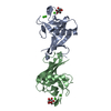
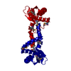
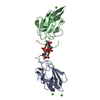
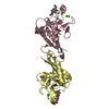
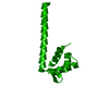
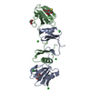
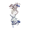
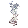
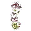
- PDB-1xsx: NMR Structure of Sso10a, a Hyperthermophile DNA-binding Protein w... -
+ Open data

- Basic information
| Entry | Database: PDB / ID: 1xsx | ||||||
|---|---|---|---|---|---|---|---|
| Title | NMR Structure of Sso10a, a Hyperthermophile DNA-binding Protein with an Extended Anti-parallel Coiled Coil | ||||||
 Components Components | Sso10a | ||||||
 Keywords Keywords | DNA BINDING PROTEIN / winged helix-turn-helix / anti-parallel coiled coil dimer / hyperthermophile DNA-binding protein | ||||||
| Function / homology |  Function and homology information Function and homology information | ||||||
| Biological species |   Sulfolobus solfataricus (archaea) Sulfolobus solfataricus (archaea) | ||||||
| Method | SOLUTION NMR / Simulated annealing using cartesion, torsion angle dynamics. | ||||||
| Model type details | minimized average | ||||||
 Authors Authors | Kahsai, M.A. / Vogler, B. / Clark, A.T. / Edmondson, S.P. / Shriver, J.W. | ||||||
 Citation Citation | Journal: Biochemistry / Year: 2005 Title: Solution Structure, Stability, and Flexibility of Sso10a: A Hyperthermophile Coiled-Coil DNA-Binding Protein(,). Authors: Kahsai, M.A. / Vogler, B. / Clark, A.T. / Edmondson, S.P. / Shriver, J.W. | ||||||
| History |
|
- Structure visualization
| Structure viewer | Molecule: MolmilJmol/JSmol |
|---|
- Downloads & links
-Download
| PDBx/mmCIF format | 1xsx.cif.gz | 756 KB | Display | PDBx/mmCIF format |
|---|---|---|---|---|
| PDB format | pdb1xsx.ent.gz | 644.3 KB | Display | PDB format |
| PDBx/mmJSON format | 1xsx.json.gz | Tree view | PDBx/mmJSON format | |
| Others |  Other downloads Other downloads |
-Validation report
| Summary document | 1xsx_validation.pdf.gz | 368.9 KB | Display | wwPDB validaton report |
|---|---|---|---|---|
| Full document | 1xsx_full_validation.pdf.gz | 557 KB | Display | |
| Data in XML | 1xsx_validation.xml.gz | 48 KB | Display | |
| Data in CIF | 1xsx_validation.cif.gz | 69.6 KB | Display | |
| Arichive directory | https://data.pdbj.org/pub/pdb/validation_reports/xs/1xsxftp://data.pdbj.org/pub/pdb/validation_reports/xs/1xsx | HTTPS FTP |
-Related structure data
| Similar structure data |
|---|

-Links
 PDBj
PDBj- Assembly
| Deposited unit | 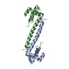
| |||||||||
|---|---|---|---|---|---|---|---|---|---|---|
| 1 |
| |||||||||
| NMR ensembles |
|
-Components
| #1: Protein | Mass: 11103.207 Da / Num. of mol.: 2 Source method: isolated from a genetically manipulated source Source: (gene. exp.) Sulfolobus solfataricus (archaea) / Gene: sso10a / Plasmid: pETBlue-2 / Production host:  |
|---|
-Experimental details
-Experiment
| Experiment | Method: SOLUTION NMR | ||||||||||||||||||||||||||||
|---|---|---|---|---|---|---|---|---|---|---|---|---|---|---|---|---|---|---|---|---|---|---|---|---|---|---|---|---|---|
| NMR experiment |
| ||||||||||||||||||||||||||||
| NMR details | Text: model 1 is an energy minimized average structure. Models 2 through 11 are the 10 best ensemble structures. |
 HSQC
HSQC- Sample preparation
| Details |
| |||||||||||||||
|---|---|---|---|---|---|---|---|---|---|---|---|---|---|---|---|---|
| Sample conditions |
|
-NMR measurement
| Radiation | Protocol: SINGLE WAVELENGTH / Monochromatic (M) / Laue (L): M | |||||||||||||||
|---|---|---|---|---|---|---|---|---|---|---|---|---|---|---|---|---|
| Radiation wavelength | Relative weight: 1 | |||||||||||||||
| NMR spectrometer |
|
- Processing
| NMR software |
| ||||||||||||||||||||||||||||||||
|---|---|---|---|---|---|---|---|---|---|---|---|---|---|---|---|---|---|---|---|---|---|---|---|---|---|---|---|---|---|---|---|---|---|
| Refinement | Method: Simulated annealing using cartesion, torsion angle dynamics. Software ordinal: 1 Details: NOE assignments, distance restraint calibrations, and initial structures were made using ARIA 1.2. Final structure refinemnet was done using Xplor-NIH. Restraints consisted of 1231 ...Details: NOE assignments, distance restraint calibrations, and initial structures were made using ARIA 1.2. Final structure refinemnet was done using Xplor-NIH. Restraints consisted of 1231 intrasubunit and 83 intersubunit NOE-derived distances, 144 torsion angles, 60 HNHA coupling constants, 41 H-bonds, 52 dipolar couplings, 54 T1/T2 relaxation ratios, and non-crystallographic dimer symmetry restraints. (Numbers of restraints are per monomer.) Anisotropy tensors were included as variables during refinemnet, with best fit values for the molecular alignment tensor (from RDC's) of Da=16 Hz and rhombicity=0.24, and best fit values for the diffusion anisotropy tensor (from 15N relaxation) of Dpar/Dperp=2.0 and rhombicity=0.05 (and tauC=13.7 ns). | ||||||||||||||||||||||||||||||||
| NMR representative | Selection criteria: minimized average structure | ||||||||||||||||||||||||||||||||
| NMR ensemble | Conformer selection criteria: 10 smallest RMSD out of 13 lowest energy Conformers calculated total number: 75 / Conformers submitted total number: 11 |
