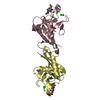
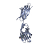
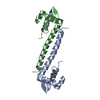
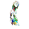
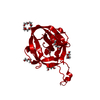
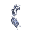
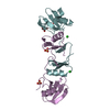
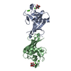
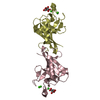
Entry Database : PDB / ID : 4wqqTitle Structure of EPNH mutant of CEL-I Lectin CEL-I, N-acetyl-D-galactosamine-specific C-type Keywords / / / / Function / homology Function Domain/homology Component
/ / / / / / / / / / / / / / / / / / / / Biological species Cucumaria echinata (invertebrata)Method / / Resolution : 1.7 Å Authors Unno, H. / Hatakeyama, T. Journal : Biochim.Biophys.Acta / Year : 2015Title : Mannose-recognition mutant of the galactose/N-acetylgalactosamine-specific C-type lectin CEL-I engineered by site-directed mutagenesis.Authors : Moriuchi, H. / Unno, H. / Goda, S. / Tateno, H. / Hirabayashi, J. / Hatakeyama, T. History Deposition Oct 22, 2014 Deposition site / Processing site Revision 1.0 Apr 29, 2015 Provider / Type Revision 1.1 May 20, 2015 Group Revision 1.2 Jul 29, 2020 Group Data collection / Database references ... Data collection / Database references / Derived calculations / Other / Source and taxonomy / Structure summary Category chem_comp / citation ... chem_comp / citation / entity / entity_src_gen / pdbx_chem_comp_identifier / pdbx_database_status / pdbx_entity_nonpoly / pdbx_struct_assembly / pdbx_struct_assembly_gen / pdbx_struct_assembly_prop / pdbx_struct_conn_angle / pdbx_struct_oper_list / struct_conn / struct_site / struct_site_gen Item _chem_comp.name / _chem_comp.type ... _chem_comp.name / _chem_comp.type / _citation.journal_id_CSD / _entity.pdbx_description / _entity_src_gen.pdbx_alt_source_flag / _pdbx_database_status.pdb_format_compatible / _pdbx_entity_nonpoly.name / _pdbx_struct_assembly.oligomeric_details / _pdbx_struct_assembly_gen.asym_id_list / _pdbx_struct_assembly_prop.type / _pdbx_struct_assembly_prop.value / _pdbx_struct_conn_angle.ptnr1_auth_seq_id / _pdbx_struct_conn_angle.ptnr3_auth_seq_id / _pdbx_struct_conn_angle.value / _pdbx_struct_oper_list.symmetry_operation / _struct_conn.pdbx_dist_value / _struct_conn.ptnr1_auth_asym_id / _struct_conn.ptnr1_auth_comp_id / _struct_conn.ptnr1_auth_seq_id / _struct_conn.ptnr1_label_asym_id / _struct_conn.ptnr1_label_atom_id / _struct_conn.ptnr1_label_comp_id / _struct_conn.ptnr1_label_seq_id / _struct_conn.ptnr1_symmetry / _struct_conn.ptnr2_auth_asym_id / _struct_conn.ptnr2_auth_comp_id / _struct_conn.ptnr2_auth_seq_id / _struct_conn.ptnr2_label_asym_id / _struct_conn.ptnr2_label_atom_id / _struct_conn.ptnr2_label_comp_id / _struct_conn.ptnr2_label_seq_id / _struct_conn.ptnr2_symmetry Description / Provider / Type Revision 1.3 Nov 8, 2023 Group Data collection / Database references ... Data collection / Database references / Refinement description / Structure summary Category chem_comp / chem_comp_atom ... chem_comp / chem_comp_atom / chem_comp_bond / database_2 / pdbx_initial_refinement_model Item / _database_2.pdbx_DOI / _database_2.pdbx_database_accessionRevision 1.4 Nov 6, 2024 Group / Category / pdbx_modification_feature
Show all Show less
 Movie
Movie Controller


 Open data
 Basic information
 Components
 Keywords
 Function and homology information
 Cucumaria echinata (invertebrata)
 X-RAY DIFFRACTION /
  Authors
 Citation
 Structure visualization
 Downloads & links
 Other downloads
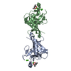


 PDBj
PDBj


 Assembly
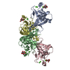


 Mass: 40.078 Da / Num. of mol.: 12 / Source method: obtained synthetically / Formula: Ca
 Type: D-saccharide, alpha linking / Mass: 180.156 Da / Num. of mol.: 4 / Source method: obtained synthetically / Formula: C6H12O6
 Mass: 18.015 Da / Num. of mol.: 758 / Source method: isolated from a natural source / Formula: H2O
 Sample preparation
 Processing