- PDB-6frg: Crystal structure of G-1F mutant of Ssp DnaB Mini-Intein variant M86 -
+
Open data
ID or keywords:
Loading...
-
Basic information
Entry
Database: PDB / ID: 6frg
Title
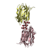
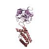
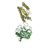
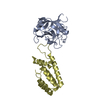
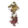
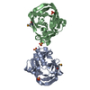
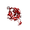
Crystal structure of G-1F mutant of Ssp DnaB Mini-Intein variant M86
Components
Replicative DNA helicase
Keywords
SPLICING / intein / protein splicing / Hint domain fold / pre-splicing form
Function / homology
Function and homology information
primosome complex / intein-mediated protein splicing / DNA replication, synthesis of primer / DNA 5'-3' helicase / DNA helicase activity / endonuclease activity / 5'-3' DNA helicase activity / Hydrolases; Acting on ester bonds / DNA replication / ATP hydrolysis activity ...primosome complex / intein-mediated protein splicing / DNA replication, synthesis of primer / DNA 5'-3' helicase / DNA helicase activity / endonuclease activity / 5'-3' DNA helicase activity / Hydrolases; Acting on ester bonds / DNA replication / ATP hydrolysis activity / DNA binding / ATP binding / cytosol Similarity search - Function
Endonuclease - Pi-scei; Chain A, domain 1 / Hedgehog/Intein (Hint) domain / DNA helicase, DnaB type / DNA helicase, DnaB-like, N-terminal / DnaB-like helicase N terminal domain / DNA helicase, DnaB-like, N-terminal domain superfamily / DNA helicase DnaB, N-terminal/DNA primase DnaG, C-terminal / LAGLIDADG-like domain / DnaB-like helicase C terminal domain / Intein splicing domain ...Endonuclease - Pi-scei; Chain A, domain 1 / Hedgehog/Intein (Hint) domain / DNA helicase, DnaB type / DNA helicase, DnaB-like, N-terminal / DnaB-like helicase N terminal domain / DNA helicase, DnaB-like, N-terminal domain superfamily / DNA helicase DnaB, N-terminal/DNA primase DnaG, C-terminal / LAGLIDADG-like domain / DnaB-like helicase C terminal domain / Intein splicing domain / DNA helicase, DnaB-like, C-terminal / Superfamily 4 helicase domain profile. / Intein / Homing endonuclease, LAGLIDADG / Intein DOD homing endonuclease / Intein DOD-type homing endonuclease domain profile. / Intein C-terminal splicing region / Intein C-terminal splicing motif profile. / Hint domain C-terminal / Hint (Hedgehog/Intein) domain C-terminal region / Homing endonuclease / Intein N-terminal splicing region / Intein N-terminal splicing motif profile. / Hint domain N-terminal / Hint (Hedgehog/Intein) domain N-terminal region / Hint domain superfamily / Beta Complex / P-loop containing nucleoside triphosphate hydrolase / Mainly Beta Similarity search - Domain/homology
In the structure databanks used in Yorodumi, some data are registered as the other names, "COVID-19 virus" and "2019-nCoV". Here are the details of the virus and the list of structure data.
Jan 31, 2019. EMDB accession codes are about to change! (news from PDBe EMDB page)
EMDB accession codes are about to change! (news from PDBe EMDB page)
The allocation of 4 digits for EMDB accession codes will soon come to an end. Whilst these codes will remain in use, new EMDB accession codes will include an additional digit and will expand incrementally as the available range of codes is exhausted. The current 4-digit format prefixed with “EMD-” (i.e. EMD-XXXX) will advance to a 5-digit format (i.e. EMD-XXXXX), and so on. It is currently estimated that the 4-digit codes will be depleted around Spring 2019, at which point the 5-digit format will come into force.
The EM Navigator/Yorodumi systems omit the EMD- prefix.
Related info.:Q: What is EMD? / ID/Accession-code notation in Yorodumi/EM Navigator
Yorodumi is a browser for structure data from EMDB, PDB, SASBDB, etc.
This page is also the successor to EM Navigator detail page, and also detail information page/front-end page for Omokage search.
The word "yorodu" (or yorozu) is an old Japanese word meaning "ten thousand". "mi" (miru) is to see.
Related info.:EMDB / PDB / SASBDB / Comparison of 3 databanks / Yorodumi Search / Aug 31, 2016. New EM Navigator & Yorodumi / Yorodumi Papers / Jmol/JSmol / Function and homology information / Changes in new EM Navigator and Yorodumi
 Movie
Movie Controller

 Yorodumi
 Open data
 Basic information
 Components
 Keywords
 Function and homology information
 X-RAY DIFFRACTION /
  Authors
 Germany, 1items
  Citation
 Structure visualization
 Downloads & links
 Other downloads


 PDBj
PDBj


 Assembly
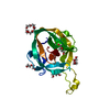

 Mass: 238.278 Da / Num. of mol.: 2 / Source method: obtained synthetically / Formula: C10H22O6 / Comment: precipitant*YM
 Mass: 150.173 Da / Num. of mol.: 1 / Source method: obtained synthetically / Formula: C6H14O4
 Mass: 106.120 Da / Num. of mol.: 4 / Source method: obtained synthetically / Formula: C4H10O3
 Mass: 18.015 Da / Num. of mol.: 221 / Source method: isolated from a natural source / Formula: H2O
 Sample preparation
 / Beamline: X06DA / Wavelength: 1 Å
 Processing