Mass: 294.400 Da / Num. of mol.: 5 / Source method: obtained synthetically / Formula: H18IrN6
-
Experimental details
-
Experiment
Experiment
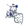
Method: X-RAY DIFFRACTION
-
Sample preparation
Crystal
Density Matthews: 2.7 Å3/Da / Density % sol: 54.4 %
Crystal grow
Temperature: 293 K / Method: vapor diffusion, hanging drop / pH: 5.6 Details: 0.1 M sodium citrate pH 5.6, 0.2 M ammonium acetate, 18% PEG 4000. Incubated with 10 mM iridium hexammine for heavy atom derivation.
Protocol: SINGLE WAVELENGTH / Monochromatic (M) / Laue (L): M / Scattering type: x-ray
Radiation wavelength
Wavelength: 1.10528 Å / Relative weight: 1
Reflection
Resolution: 3.1→20 Å / Num. obs: 9586 / % possible obs: 98.9 % / Redundancy: 7.6 % / Rmerge(I) obs: 0.049 / Net I/σ(I): 26.7
Reflection shell
Resolution: 3.1→3.18 Å / Redundancy: 7.7 % / Rmerge(I) obs: 0.242 / Mean I/σ(I) obs: 7.45 / Num. unique all: 677 / % possible all: 98.8
-
Processing
Software
Name
Version
Classification
XDS
datareduction
XSCALE
datascaling
PHENIX
phasing
Coot
modelbuilding
PHENIX
1.9_1692
refinement
Refinement
Method to determine structure: SAD / Resolution: 3.1003→19.355 Å / SU ML: 0.37 / Cross valid method: THROUGHOUT / σ(F): 1.36 / Phase error: 21.85 / Stereochemistry target values: ML
Rfactor
Num. reflection
% reflection
Rfree
0.2273
243
4.56 %
Rwork
0.2007
-
-
obs
0.2019
5334
98.92 %
Solvent computation
Shrinkage radii: 0.9 Å / VDW probe radii: 1.11 Å / Solvent model: FLAT BULK SOLVENT MODEL
Refinement step
Cycle: LAST / Resolution: 3.1003→19.355 Å
Protein
Nucleic acid
Ligand
Solvent
Total
Num. atoms
170
1518
37
0
1725
Refine LS restraints
Refine-ID
Type
Dev ideal
Number
X-RAY DIFFRACTION
f_bond_d
0.006
1898
X-RAY DIFFRACTION
f_angle_d
1.217
2943
X-RAY DIFFRACTION
f_dihedral_angle_d
18.709
896
X-RAY DIFFRACTION
f_chiral_restr
0.058
360
X-RAY DIFFRACTION
f_plane_restr
0.006
104
LS refinement shell
Resolution (Å)
Rfactor Rfree
Num. reflection Rfree
Rfactor Rwork
Num. reflection Rwork
Refine-ID
% reflection obs (%)
3.1003-3.9008
0.3
131
0.2423
2437
X-RAY DIFFRACTION
98
3.9008-19.3558
0.1713
112
0.173
2654
X-RAY DIFFRACTION
100
Refinement TLS params.
Method: refined / Refine-ID: X-RAY DIFFRACTION
ID
L11 (°2)
L12 (°2)
L13 (°2)
L22 (°2)
L23 (°2)
L33 (°2)
S11 (Å °)
S12 (Å °)
S13 (Å °)
S21 (Å °)
S22 (Å °)
S23 (Å °)
S31 (Å °)
S32 (Å °)
S33 (Å °)
T11 (Å2)
T12 (Å2)
T13 (Å2)
T22 (Å2)
T23 (Å2)
T33 (Å2)
Origin x (Å)
Origin y (Å)
Origin z (Å)
1
0.5757
0.1602
-0.0205
0.6787
-0.0426
1.7809
0.0938
-0.2115
-0.3011
0.118
-0.0619
0.214
-0.186
-0.4444
0.4027
0.1081
-0.0165
-0.0591
0.2723
0.0565
0.3933
-12.3668
38.8447
13.1761
2
3.9012
-0.2865
1.0116
3.7887
-1.9621
1.4082
-0.0727
0.8046
0.2462
-1.1424
0.065
-0.7247
0.2475
0.17
0.18
0.3229
-0.1418
0.102
0.3754
0.04
0.2877
-10.0495
34.665
1.5231
3
3.3763
3.1341
-1.0551
3.3019
-1.9291
2.6532
0.2992
-0.2284
0.7038
0.6258
-0.2052
1.2212
-1.1212
-0.4787
-0.1161
0.4799
0.1929
0.0293
0.3269
0.0817
0.534
-15.9132
51.7617
11.0398
4
2.6932
0.252
-0.3609
3.2699
-0.2601
0.5201
0.1019
0.3973
0.4536
-0.6689
0.1804
-0.0591
-0.4505
-0.0746
0.1088
0.4481
-0.0238
0.0131
0.0935
0.0513
0.2824
-9.3434
49.1259
6.3682
5
0.5977
0.7556
-0.4334
1.0296
-0.5444
0.3238
-0.1766
-0.0273
-0.2071
0.4211
-0.3276
-0.0452
0.0292
-0.0091
-0.7772
0.7484
0.0142
0.0245
0.1427
-0.1873
0.2956
-10.3295
74.8369
-3.6608
6
2.2
-0.72
-1.7232
4.1429
-0.5812
1.6795
0.3102
-0.0069
-0.2832
-0.1459
-0.5107
0.5695
-0.2137
0.1009
-0.4658
0.9832
-0.0053
-0.0801
0.085
-0.0171
0.2481
-15.3289
87.6409
-5.971
7
5.1608
0.2221
1.4071
0.1777
0.0081
0.4029
-0.0316
-0.8272
0.4397
0.8363
-0.173
0.8184
0.1786
-0.2838
0.3424
1.0173
0.1178
0.346
0.4397
-0.0229
0.6761
-22.3003
86.1316
3.6512
8
1.6568
-0.6262
0.5202
2.3234
0.3771
0.7483
0.0783
-0.0364
-0.4446
0.5284
-0.2381
0.8095
-0.2547
-0.0683
0.1031
0.9205
0.1542
0.0661
0.2357
-0.12
0.4621
-13.9202
71.4636
-2.0908
9
2.3079
-0.239
1.2795
4.8958
-1.2075
0.9446
0.3589
-0.1255
-0.4864
0.5152
-0.1288
0.4931
-0.2505
-0.2701
-0.0074
0.8078
0.0201
0.043
0.2805
-0.0764
0.3762
-12.8276
73.2559
3.4173
Refinement TLS group
ID
Refine-ID
Refine TLS-ID
Selection details
1
X-RAY DIFFRACTION
1
chain 'A' and (resid1through21 )
2
X-RAY DIFFRACTION
2
chain 'A' and (resid22through28 )
3
X-RAY DIFFRACTION
3
chain 'A' and (resid29through35 )
4
X-RAY DIFFRACTION
4
chain 'B' and (resid5through18 )
5
X-RAY DIFFRACTION
5
chain 'C' and (resid1through9 )
6
X-RAY DIFFRACTION
6
chain 'C' and (resid10through21 )
7
X-RAY DIFFRACTION
7
chain 'C' and (resid22through26 )
8
X-RAY DIFFRACTION
8
chain 'C' and (resid27through35 )
9
X-RAY DIFFRACTION
9
chain 'D' and (resid6through19 )
+
About Yorodumi
-
News
-
Feb 9, 2022. New format data for meta-information of EMDB entries
New format data for meta-information of EMDB entries
Version 3 of the EMDB header file is now the official format.
The previous official version 1.9 will be removed from the archive.
In the structure databanks used in Yorodumi, some data are registered as the other names, "COVID-19 virus" and "2019-nCoV". Here are the details of the virus and the list of structure data.
Jan 31, 2019. EMDB accession codes are about to change! (news from PDBe EMDB page)
EMDB accession codes are about to change! (news from PDBe EMDB page)
The allocation of 4 digits for EMDB accession codes will soon come to an end. Whilst these codes will remain in use, new EMDB accession codes will include an additional digit and will expand incrementally as the available range of codes is exhausted. The current 4-digit format prefixed with “EMD-” (i.e. EMD-XXXX) will advance to a 5-digit format (i.e. EMD-XXXXX), and so on. It is currently estimated that the 4-digit codes will be depleted around Spring 2019, at which point the 5-digit format will come into force.
The EM Navigator/Yorodumi systems omit the EMD- prefix.
Related info.:Q: What is EMD? / ID/Accession-code notation in Yorodumi/EM Navigator
Yorodumi is a browser for structure data from EMDB, PDB, SASBDB, etc.
This page is also the successor to EM Navigator detail page, and also detail information page/front-end page for Omokage search.
The word "yorodu" (or yorozu) is an old Japanese word meaning "ten thousand". "mi" (miru) is to see.
Related info.:EMDB / PDB / SASBDB / Comparison of 3 databanks / Yorodumi Search / Aug 31, 2016. New EM Navigator & Yorodumi / Yorodumi Papers / Jmol/JSmol / Function and homology information / Changes in new EM Navigator and Yorodumi
 Movie
Movie Controller

 Yorodumi
 Open data
 Basic information
 Components
 Keywords
 Function and homology information
 Homo sapiens (human)
 X-RAY DIFFRACTION /
  Authors
 United States, 6items
  Citation
 Structure visualization
 Downloads & links
 Other downloads


 PDBj
PDBj


 Assembly
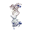


 Mass: 39.098 Da / Num. of mol.: 2 / Source method: obtained synthetically / Formula: K
 Mass: 294.400 Da / Num. of mol.: 5 / Source method: obtained synthetically / Formula: H18IrN6
 Sample preparation
 Processing