 Movie
Movie Controller

[English] 日本語
 Yorodumi
- PDB-2yip: Crystal Structure of Parasite Sarcocystis muris Microneme Protein... -
+ Open data

- Basic information
| Entry | Database: PDB / ID: 2yip | ||||||
|---|---|---|---|---|---|---|---|
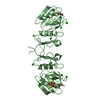
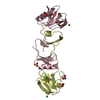
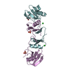
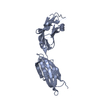
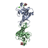
| Title | Crystal Structure of Parasite Sarcocystis muris Microneme Protein SML- 2 in complex with 1-Thio-beta-D-Galactose (SPACEGROUP P212121) | ||||||
 Components Components | MICRONEME ANTIGEN L2 | ||||||
 Keywords Keywords | SUGAR BINDING PROTEIN / APPLE-DOMAIN TANDEM REPEAT / PAN_AP / PAN_1 / GALACTOSE-BINDING LECTIN / CELLULAR ADHESION / MICRONEMAL PROTEIN | ||||||
| Function / homology |  Function and homology information Function and homology informationmicroneme / carbohydrate binding / cytoplasmic vesicle / cell adhesion / proteolysis / extracellular region Similarity search - Function | ||||||
| Biological species |  SARCOCYSTIS MURIS (eukaryote) SARCOCYSTIS MURIS (eukaryote) | ||||||
| Method |  X-RAY DIFFRACTION / SYNCHROTRON / MOLECULAR REPLACEMENT / Resolution: 2.14 Å X-RAY DIFFRACTION / SYNCHROTRON / MOLECULAR REPLACEMENT / Resolution: 2.14 Å | ||||||
 Authors Authors | Mueller, J.J. / Heinemann, U. | ||||||
 Citation Citation | Journal: Acta Crystallogr.,Sect.D / Year: 2011 Title: Pan-Modular Structure of Microneme Protein Sml-2 from Parasite Sarcocystis Muris at 1.95 A Resolution and its Complex with 1-Thio-Beta-D-Galactose. Authors: Mueller, J.J. / Weiss, M.S. / Heinemann, U. | ||||||
| History |
|
- Structure visualization
| Structure viewer | Molecule: MolmilJmol/JSmol |
|---|
- Downloads & links
-Download
| PDBx/mmCIF format | 2yip.cif.gz | 172.7 KB | Display | PDBx/mmCIF format |
|---|---|---|---|---|
| PDB format | pdb2yip.ent.gz | 138.8 KB | Display | PDB format |
| PDBx/mmJSON format | 2yip.json.gz | Tree view | PDBx/mmJSON format | |
| Others |  Other downloads Other downloads |
-Validation report
| Arichive directory | https://data.pdbj.org/pub/pdb/validation_reports/yi/2yipftp://data.pdbj.org/pub/pdb/validation_reports/yi/2yip | HTTPS FTP |
|---|
-Related structure data
| Related structure data |  2yilSC 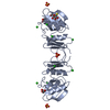 2yioC S: Starting model for refinement C: citing same article ( |
|---|---|
| Similar structure data |

-Links
 PDBj
PDBj- Assembly
| Deposited unit | 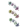
| |||||||||||||||||||||||||||||||||||||||||||||||||||||||||||||||||||||
|---|---|---|---|---|---|---|---|---|---|---|---|---|---|---|---|---|---|---|---|---|---|---|---|---|---|---|---|---|---|---|---|---|---|---|---|---|---|---|---|---|---|---|---|---|---|---|---|---|---|---|---|---|---|---|---|---|---|---|---|---|---|---|---|---|---|---|---|---|---|---|
| 1 | 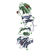
| |||||||||||||||||||||||||||||||||||||||||||||||||||||||||||||||||||||
| 2 | 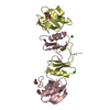
| |||||||||||||||||||||||||||||||||||||||||||||||||||||||||||||||||||||
| 3 | 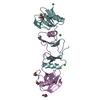
| |||||||||||||||||||||||||||||||||||||||||||||||||||||||||||||||||||||
| Unit cell |
| |||||||||||||||||||||||||||||||||||||||||||||||||||||||||||||||||||||
| Noncrystallographic symmetry (NCS) | NCS domain:
NCS domain segments: Component-ID: 1 / Beg auth comp-ID: VAL / Beg label comp-ID: VAL / End auth comp-ID: TYR / End label comp-ID: TYR / Refine code: 4 / Auth seq-ID: 7 - 130 / Label seq-ID: 7 - 130
NCS ensembles :
NCS oper:
|
-Components
| #1: Protein | Mass: 15094.004 Da / Num. of mol.: 6 / Source method: isolated from a natural source Details: CYST MEROZOITES OF SARCOCYSTIS MURIS FROM SKELETAL MOUSE MUSCLES. Source: (natural) SARCOCYSTIS MURIS (eukaryote) / References: UniProt: P81860#2: Sugar | ChemComp-YIO /   Type: D-saccharide, beta linking / Mass: 196.221 Da / Num. of mol.: 6 / Source method: obtained synthetically / Formula: C6H12O5S Type: D-saccharide, beta linking / Mass: 196.221 Da / Num. of mol.: 6 / Source method: obtained synthetically / Formula: C6H12O5S#3: Chemical | ChemComp-CL /   Mass: 35.453 Da / Num. of mol.: 13 / Source method: obtained synthetically / Formula: Cl Mass: 35.453 Da / Num. of mol.: 13 / Source method: obtained synthetically / Formula: Cl#4: Chemical | ChemComp-GOL /   Mass: 92.094 Da / Num. of mol.: 7 / Source method: obtained synthetically / Formula: C3H8O3 Mass: 92.094 Da / Num. of mol.: 7 / Source method: obtained synthetically / Formula: C3H8O3#5: Water | ChemComp-HOH / |  Mass: 18.015 Da / Num. of mol.: 517 / Source method: isolated from a natural source / Formula: H2O Mass: 18.015 Da / Num. of mol.: 517 / Source method: isolated from a natural source / Formula: H2OHas protein modification | Y | |
|---|
-Experimental details
-Experiment
| Experiment | Method: X-RAY DIFFRACTION / Number of used crystals: 1 |
|---|
- Sample preparation
| Crystal | Density Matthews: 3.06 Å3/Da / Density % sol: 59.8 % / Description: NONE |
|---|---|
| Crystal grow | Temperature: 291 K / Method: vapor diffusion, hanging drop / pH: 7.9 Details: VAPOR DIFFUSION, HANGING DROP. PH 7.9, TEMPERATURE 291K. VAPOR DIFFUSION, HANGING DROP. PROTEIN SOLUTION: 50 MM TRIS-HCL, 150 MM NACL, PH 8.3, 12MG/ML SML-2. RESERVOIR: 0.1 M HEPES, PH 7.5, ...Details: VAPOR DIFFUSION, HANGING DROP. PH 7.9, TEMPERATURE 291K. VAPOR DIFFUSION, HANGING DROP. PROTEIN SOLUTION: 50 MM TRIS-HCL, 150 MM NACL, PH 8.3, 12MG/ML SML-2. RESERVOIR: 0.1 M HEPES, PH 7.5, 0.1 M NACL, 1.7 M AMMONIUM SULFATE, 15% GLYCEROL. DROPLET: 1 MICROLITER PROTEIN SOLUTION: 1 MICROLITER RESERVOIR SOLUTION. PROTEIN:1-THIO-BETA-D-GALACTOSE MOLAR RATIO 1:100. |
-Data collection
| Diffraction | Mean temperature: 110 K |
|---|---|
| Diffraction source | Source: SYNCHROTRON / Site: EMBL/DESY, HAMBURG  / Beamline: BW7B / Wavelength: 0.8428 / Beamline: BW7B / Wavelength: 0.8428 |
| Detector | Type: MAR scanner 345 mm plate / Detector: IMAGE PLATE / Date: May 19, 2000 / Details: MIRRORS |
| Radiation | Monochromator: SI(111) / Protocol: SINGLE WAVELENGTH / Monochromatic (M) / Laue (L): M / Scattering type: x-ray |
| Radiation wavelength | Wavelength: 0.8428 Å / Relative weight: 1 |
| Reflection | Resolution: 2.14→35 Å / Num. obs: 61346 / % possible obs: 99.5 % / Observed criterion σ(I): -3 / Redundancy: 5 % / Biso Wilson estimate: 28.6 Å2 / Rmerge(I) obs: 0.03 / Net I/σ(I): 38.6 |
| Reflection shell | Resolution: 2.14→2.2 Å / Redundancy: 4.6 % / Rmerge(I) obs: 0.1 / Mean I/σ(I) obs: 14.1 / % possible all: 95.8 |
- Processing
| Software |
| ||||||||||||||||||||||||||||||||||||||||||||||||||||||||||||||||||||||||||||||||||||||||||||||||||||||||||||||||||||||||||||||||||||||||||||||||||||||||||||||||||||||||||||||||||||||
|---|---|---|---|---|---|---|---|---|---|---|---|---|---|---|---|---|---|---|---|---|---|---|---|---|---|---|---|---|---|---|---|---|---|---|---|---|---|---|---|---|---|---|---|---|---|---|---|---|---|---|---|---|---|---|---|---|---|---|---|---|---|---|---|---|---|---|---|---|---|---|---|---|---|---|---|---|---|---|---|---|---|---|---|---|---|---|---|---|---|---|---|---|---|---|---|---|---|---|---|---|---|---|---|---|---|---|---|---|---|---|---|---|---|---|---|---|---|---|---|---|---|---|---|---|---|---|---|---|---|---|---|---|---|---|---|---|---|---|---|---|---|---|---|---|---|---|---|---|---|---|---|---|---|---|---|---|---|---|---|---|---|---|---|---|---|---|---|---|---|---|---|---|---|---|---|---|---|---|---|---|---|---|---|
| Refinement | Method to determine structure: MOLECULAR REPLACEMENT Starting model: PDB ENTRY 2YIL Resolution: 2.14→33.89 Å / Cor.coef. Fo:Fc: 0.958 / Cor.coef. Fo:Fc free: 0.936 / SU B: 3.402 / SU ML: 0.091 / Cross valid method: THROUGHOUT / ESU R: 0.158 / ESU R Free: 0.149 / Stereochemistry target values: MAXIMUM LIKELIHOOD Details: HYDROGENS HAVE BEEN ADDED IN THE RIDING POSITIONS. U VALUES REFINED INDIVIDUALLY.
| ||||||||||||||||||||||||||||||||||||||||||||||||||||||||||||||||||||||||||||||||||||||||||||||||||||||||||||||||||||||||||||||||||||||||||||||||||||||||||||||||||||||||||||||||||||||
| Solvent computation | Ion probe radii: 0.8 Å / Shrinkage radii: 0.8 Å / VDW probe radii: 1.4 Å / Solvent model: BABINET MODEL WITH MASK | ||||||||||||||||||||||||||||||||||||||||||||||||||||||||||||||||||||||||||||||||||||||||||||||||||||||||||||||||||||||||||||||||||||||||||||||||||||||||||||||||||||||||||||||||||||||
| Displacement parameters | Biso mean: 29.115 Å2
| ||||||||||||||||||||||||||||||||||||||||||||||||||||||||||||||||||||||||||||||||||||||||||||||||||||||||||||||||||||||||||||||||||||||||||||||||||||||||||||||||||||||||||||||||||||||
| Refinement step | Cycle: LAST / Resolution: 2.14→33.89 Å
| ||||||||||||||||||||||||||||||||||||||||||||||||||||||||||||||||||||||||||||||||||||||||||||||||||||||||||||||||||||||||||||||||||||||||||||||||||||||||||||||||||||||||||||||||||||||
| Refine LS restraints |
|