- PDB-1xk8: Divalent cation tolerant protein CUTA from Homo sapiens O60888 -
+
データを開く
IDまたはキーワード:
読み込み中...
-
基本情報
登録情報
データベース: PDB / ID: 1xk8
タイトル
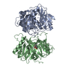
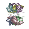
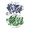
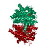
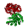
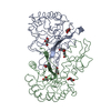
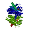
Divalent cation tolerant protein CUTA from Homo sapiens O60888
要素
Divalent cation tolerant protein CUTA
キーワード
structural genomics / unknown function / human / Homo sapiens / divalent cation tolerant protein CUTA / PSI / Protein Structure Initiative / Southeast Collaboratory for Structural Genomics / SECSG
機能・相同性
機能・相同性情報
response to metal ion / intracellular protein localization / copper ion binding / enzyme binding / mitochondrion / extracellular exosome / membrane 類似検索 - 分子機能
Divalent ion tolerance protein, CutA / CutA1 divalent ion tolerance protein / Alpha-Beta Plaits - #120 / Nitrogen regulatory PII-like, alpha/beta / Nitrogen regulatory protein PII/ATP phosphoribosyltransferase, C-terminal / Alpha-Beta Plaits / 2-Layer Sandwich / Alpha Beta 類似検索 - ドメイン・相同性
BIOMOLECULE: THIS ENTRY CONTAINS THE CRYSTALLOGRAPHIC ASYMMETRIC UNIT WHICH CONSISTS OF 6 CHAIN(S). ...BIOMOLECULE: THIS ENTRY CONTAINS THE CRYSTALLOGRAPHIC ASYMMETRIC UNIT WHICH CONSISTS OF 6 CHAIN(S). THE BIOLOGICAL UNIT IS UNKNOWN.
 ムービー
ムービー コントローラー


 データを開く
 基本情報
基本情報 要素
要素 キーワード
キーワード 機能・相同性情報
 Homo sapiens (ヒト)
 X線回折 /
  データ登録者
 引用
引用 構造の表示
構造の表示 ダウンロードとリンク
 その他のダウンロード


 PDBj
PDBj 集合体
集合体