 Movie
Movie Controller

[English] 日本語
 Yorodumi
- PDB-2zfh: Crystal structure of putative CutA1 from Homo sapiens at 2.05A re... -
+ Open data

- Basic information
| Entry | Database: PDB / ID: 2zfh | ||||||
|---|---|---|---|---|---|---|---|
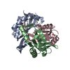
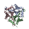
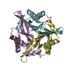
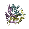
| Title | Crystal structure of putative CutA1 from Homo sapiens at 2.05A resolution | ||||||
 Components Components | CutA | ||||||
 Keywords Keywords | STRUCTURAL GENOMICS / UNKNOWN FUNCTION / Human brain / Trimeric structure / NPPSFA / National Project on Protein Structural and Functional Analyses / RIKEN Structural Genomics/Proteomics Initiative / RSGI | ||||||
| Function / homology |  Function and homology information Function and homology informationresponse to metal ion / intracellular protein localization / copper ion binding / enzyme binding / mitochondrion / extracellular exosome / membrane Similarity search - Function | ||||||
| Biological species |  Homo sapiens (human) Homo sapiens (human) | ||||||
| Method |  X-RAY DIFFRACTION / SYNCHROTRON / MOLECULAR REPLACEMENT / Resolution: 2.05 Å X-RAY DIFFRACTION / SYNCHROTRON / MOLECULAR REPLACEMENT / Resolution: 2.05 Å | ||||||
 Authors Authors | Bagautdinov, B. / Yutani, K. / RIKEN Structural Genomics/Proteomics Initiative (RSGI) | ||||||
 Citation Citation | Journal: Acta Crystallogr.,Sect.F / Year: 2008 Title: Structure of putative CutA1 from Homo sapiens determined at 2.05 A resolution. Authors: Bagautdinov, B. / Matsuura, Y. / Bagautdinova, S. / Kunishima, N. / Yutani, K. | ||||||
| History |
|
- Structure visualization
| Structure viewer | Molecule: MolmilJmol/JSmol |
|---|
- Downloads & links
-Download
| PDBx/mmCIF format | 2zfh.cif.gz | 143 KB | Display | PDBx/mmCIF format |
|---|---|---|---|---|
| PDB format | pdb2zfh.ent.gz | 110.6 KB | Display | PDB format |
| PDBx/mmJSON format | 2zfh.json.gz | Tree view | PDBx/mmJSON format | |
| Others |  Other downloads Other downloads |
-Validation report
| Arichive directory | https://data.pdbj.org/pub/pdb/validation_reports/zf/2zfhftp://data.pdbj.org/pub/pdb/validation_reports/zf/2zfh | HTTPS FTP |
|---|
-Related structure data
| Related structure data |  1xk8S S: Starting model for refinement |
|---|---|
| Similar structure data |


-Links
 PDBj
PDBj- Assembly
| Deposited unit | 
| ||||||||
|---|---|---|---|---|---|---|---|---|---|
| 1 | 
| ||||||||
| 2 | 
| ||||||||
| Unit cell |
|
-Components
| #1: Protein | Mass: 19130.230 Da / Num. of mol.: 6 Source method: isolated from a genetically manipulated source Source: (gene. exp.) Homo sapiens (human) / Gene: CUTA, ACHAP, C6orf82 / Plasmid: pET 11a / Production host:  #2: Water | ChemComp-HOH / |  Mass: 18.015 Da / Num. of mol.: 252 / Source method: isolated from a natural source / Formula: H2O Mass: 18.015 Da / Num. of mol.: 252 / Source method: isolated from a natural source / Formula: H2O |
|---|
-Experimental details
-Experiment
| Experiment | Method: X-RAY DIFFRACTION / Number of used crystals: 1 |
|---|
- Sample preparation
| Crystal | Density Matthews: 2.2 Å3/Da / Density % sol: 43 % |
|---|---|
| Crystal grow | Temperature: 295 K / Method: microbatch / pH: 4.6 Details: 25% PEG4000, 0.17M Ammonium Acetate, 0.085M Sodium acetate, 15% glycerol, pH4.6, microbatch, temperature 295K |
-Data collection
| Diffraction | Mean temperature: 100 K |
|---|---|
| Diffraction source | Source: SYNCHROTRON / Site: SPring-8  / Beamline: BL26B1 / Wavelength: 1 Å / Beamline: BL26B1 / Wavelength: 1 Å |
| Detector | Type: MARRESEARCH / Detector: CCD / Date: Jun 22, 2007 / Details: mirrors |
| Radiation | Monochromator: GRAPHITE / Protocol: SINGLE WAVELENGTH / Monochromatic (M) / Laue (L): M / Scattering type: x-ray |
| Radiation wavelength | Wavelength: 1 Å / Relative weight: 1 |
| Reflection | Resolution: 2.05→40 Å / Num. all: 48457 / Num. obs: 45947 / % possible obs: 94.8 % / Observed criterion σ(F): 0 / Observed criterion σ(I): 0 / Redundancy: 6.6 % / Biso Wilson estimate: 33.8 Å2 / Rmerge(I) obs: 0.069 / Rsym value: 0.057 / Net I/σ(I): 12.5 |
| Reflection shell | Resolution: 2.05→2.12 Å / Redundancy: 5.8 % / Rmerge(I) obs: 0.374 / Mean I/σ(I) obs: 2.3 / Num. unique all: 3214 / Rsym value: 0.335 / % possible all: 79.2 |
- Processing
| Software |
| |||||||||||||||||||||||||
|---|---|---|---|---|---|---|---|---|---|---|---|---|---|---|---|---|---|---|---|---|---|---|---|---|---|---|
| Refinement | Method to determine structure: MOLECULAR REPLACEMENT Starting model: PDB ENTRY 1XK8 Resolution: 2.05→37.81 Å / Isotropic thermal model: Overall / Cross valid method: THROUGHOUT / σ(F): 0 / σ(I): 0 / Stereochemistry target values: Engh & Huber
| |||||||||||||||||||||||||
| Displacement parameters | Biso mean: 51.1 Å2
| |||||||||||||||||||||||||
| Refine analyze |
| |||||||||||||||||||||||||
| Refinement step | Cycle: LAST / Resolution: 2.05→37.81 Å
| |||||||||||||||||||||||||
| Refine LS restraints |
|
