 Movie
Movie Controller

[English] 日本語
 Yorodumi
- PDB-1rqj: Active Conformation of Farnesyl Pyrophosphate Synthase Bound to I... -
+ Open data

- Basic information
| Entry | Database: PDB / ID: 1rqj | ||||||
|---|---|---|---|---|---|---|---|
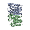
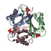
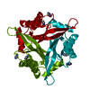
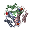
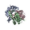
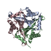
| Title | Active Conformation of Farnesyl Pyrophosphate Synthase Bound to Isopentyl Pyrophosphate and Risedronate | ||||||
 Components Components | Geranyltranstransferase | ||||||
 Keywords Keywords | TRANSFERASE / bisphosphonate / isoprenyl synthase | ||||||
| Function / homology |  Function and homology information Function and homology informationgeranyl diphosphate biosynthetic process / (2E,6E)-farnesyl diphosphate synthase / prenyltransferase activity / trans, trans-farnesyl diphosphate biosynthetic process / dimethylallyltranstransferase activity / (2E,6E)-farnesyl diphosphate synthase activity / metal ion binding / cytosol Similarity search - Function | ||||||
| Biological species |  | ||||||
| Method |  X-RAY DIFFRACTION / SYNCHROTRON / MOLECULAR REPLACEMENT / Resolution: 1.95 Å X-RAY DIFFRACTION / SYNCHROTRON / MOLECULAR REPLACEMENT / Resolution: 1.95 Å | ||||||
 Authors Authors | Hosfield, D.J. / Zhang, Y. / Dougan, D.R. / Brooun, A. / Tari, L.W. / Swanson, R.V. / Finn, J. | ||||||
 Citation Citation | Journal: J.Biol.Chem. / Year: 2004 Title: Structural basis for bisphosphonate-mediated inhibition of isoprenoid biosynthesis Authors: Hosfield, D.J. / Zhang, Y. / Dougan, D.R. / Brooun, A. / Tari, L.W. / Swanson, R.V. / Finn, J. | ||||||
| History |
|
- Structure visualization
| Structure viewer | Molecule: MolmilJmol/JSmol |
|---|
- Downloads & links
-Download
| PDBx/mmCIF format | 1rqj.cif.gz | 136 KB | Display | PDBx/mmCIF format |
|---|---|---|---|---|
| PDB format | pdb1rqj.ent.gz | 106.2 KB | Display | PDB format |
| PDBx/mmJSON format | 1rqj.json.gz | Tree view | PDBx/mmJSON format | |
| Others |  Other downloads Other downloads |
-Validation report
| Arichive directory | https://data.pdbj.org/pub/pdb/validation_reports/rq/1rqjftp://data.pdbj.org/pub/pdb/validation_reports/rq/1rqj | HTTPS FTP |
|---|
-Related structure data


-Links
 PDBj
PDBj
- Assembly
| Deposited unit | 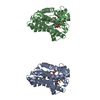
| ||||||||
|---|---|---|---|---|---|---|---|---|---|
| 1 | 
| ||||||||
| 2 | 
| ||||||||
| 3 | 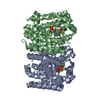
| ||||||||
| Unit cell |
|
-Components
| #1: Protein | Mass: 32197.615 Da / Num. of mol.: 2 Source method: isolated from a genetically manipulated source Source: (gene. exp.) References: UniProt: P22939, (2E,6E)-farnesyl diphosphate synthase #2: Chemical | ChemComp-MG /   Mass: 24.305 Da / Num. of mol.: 6 / Source method: obtained synthetically / Formula: Mg Mass: 24.305 Da / Num. of mol.: 6 / Source method: obtained synthetically / Formula: Mg#3: Chemical |   Mass: 246.092 Da / Num. of mol.: 2 / Source method: obtained synthetically / Formula: C5H12O7P2 Mass: 246.092 Da / Num. of mol.: 2 / Source method: obtained synthetically / Formula: C5H12O7P2#4: Chemical |   Mass: 283.112 Da / Num. of mol.: 2 / Source method: obtained synthetically / Formula: C7H11NO7P2 Mass: 283.112 Da / Num. of mol.: 2 / Source method: obtained synthetically / Formula: C7H11NO7P2#5: Water | ChemComp-HOH / |  Mass: 18.015 Da / Num. of mol.: 413 / Source method: isolated from a natural source / Formula: H2O Mass: 18.015 Da / Num. of mol.: 413 / Source method: isolated from a natural source / Formula: H2O |
|---|
-Experimental details
-Experiment
| Experiment | Method: X-RAY DIFFRACTION / Number of used crystals: 1 |
|---|
- Sample preparation
| Crystal | Density Matthews: 2.68 Å3/Da / Density % sol: 54.06 % |
|---|---|
| Crystal grow | Temperature: 298 K / Method: nanoliter sitting drop vapor diffusion / pH: 6.5 Details: MPEG 2000, HEPES, pH 6.5, Nanoliter Sitting Drop Vapour Diffusion, temperature 298K |
| Crystal grow | *PLUS Method: unknown / Details: Hosfield, D., (2003) J. Struct. Biol., 142, 207. |
-Data collection
| Diffraction | Mean temperature: 97 K |
|---|---|
| Diffraction source | Source: SYNCHROTRON / Site: ALS  / Beamline: 5.0.3 / Wavelength: 1 Å / Beamline: 5.0.3 / Wavelength: 1 Å |
| Detector | Type: ADSC QUANTUM 4 / Detector: CCD / Date: Nov 11, 2002 |
| Radiation | Protocol: SINGLE WAVELENGTH / Monochromatic (M) / Laue (L): M / Scattering type: x-ray |
| Radiation wavelength | Wavelength: 1 Å / Relative weight: 1 |
| Reflection | Resolution: 1.95→50 Å / Num. all: 52067 / Num. obs: 51703 / % possible obs: 99.3 % / Observed criterion σ(F): 2 / Observed criterion σ(I): 2 / Redundancy: 7.66 % / Rsym value: 0.077 / Net I/σ(I): 27.8 |
| Reflection shell | Resolution: 1.95→2.02 Å / Redundancy: 5.5 % / Mean I/σ(I) obs: 3 / Rsym value: 0.51 / % possible all: 96.2 |
| Reflection | *PLUS Lowest resolution: 50 Å / Num. measured all: 396530 / Rmerge(I) obs: 0.077 |
| Reflection shell | *PLUS % possible obs: 96.2 % / Rmerge(I) obs: 0.516 / Mean I/σ(I) obs: 3 |
- Processing
| Software |
| |||||||||||||||||||||||||||||||||||||||||||||||||||||||||||||||||||||||||||||||||||||||||||||||||||||||||
|---|---|---|---|---|---|---|---|---|---|---|---|---|---|---|---|---|---|---|---|---|---|---|---|---|---|---|---|---|---|---|---|---|---|---|---|---|---|---|---|---|---|---|---|---|---|---|---|---|---|---|---|---|---|---|---|---|---|---|---|---|---|---|---|---|---|---|---|---|---|---|---|---|---|---|---|---|---|---|---|---|---|---|---|---|---|---|---|---|---|---|---|---|---|---|---|---|---|---|---|---|---|---|---|---|---|---|
| Refinement | Method to determine structure: MOLECULAR REPLACEMENT / Resolution: 1.95→20 Å / Cor.coef. Fo:Fc: 0.939 / Cor.coef. Fo:Fc free: 0.923 / SU B: 3.645 / SU ML: 0.105 / Cross valid method: THROUGHOUT / ESU R: 0.17 / ESU R Free: 0.152 / Stereochemistry target values: MAXIMUM LIKELIHOOD / Details: HYDROGENS HAVE BEEN ADDED IN THE RIDING POSITIONS
| |||||||||||||||||||||||||||||||||||||||||||||||||||||||||||||||||||||||||||||||||||||||||||||||||||||||||
| Solvent computation | Ion probe radii: 0.8 Å / Shrinkage radii: 0.8 Å / VDW probe radii: 1.4 Å / Solvent model: BABINET MODEL WITH MASK | |||||||||||||||||||||||||||||||||||||||||||||||||||||||||||||||||||||||||||||||||||||||||||||||||||||||||
| Displacement parameters | Biso mean: 22.376 Å2
| |||||||||||||||||||||||||||||||||||||||||||||||||||||||||||||||||||||||||||||||||||||||||||||||||||||||||
| Refinement step | Cycle: LAST / Resolution: 1.95→20 Å
| |||||||||||||||||||||||||||||||||||||||||||||||||||||||||||||||||||||||||||||||||||||||||||||||||||||||||
| Refine LS restraints |
| |||||||||||||||||||||||||||||||||||||||||||||||||||||||||||||||||||||||||||||||||||||||||||||||||||||||||
| LS refinement shell | Resolution: 1.95→1.998 Å / Total num. of bins used: 20 /
| |||||||||||||||||||||||||||||||||||||||||||||||||||||||||||||||||||||||||||||||||||||||||||||||||||||||||
| Refinement | *PLUS Lowest resolution: 20 Å / % reflection Rfree: 5 % / Rfactor Rfree: 0.24 / Rfactor Rwork: 0.2 | |||||||||||||||||||||||||||||||||||||||||||||||||||||||||||||||||||||||||||||||||||||||||||||||||||||||||
| Solvent computation | *PLUS | |||||||||||||||||||||||||||||||||||||||||||||||||||||||||||||||||||||||||||||||||||||||||||||||||||||||||
| Displacement parameters | *PLUS | |||||||||||||||||||||||||||||||||||||||||||||||||||||||||||||||||||||||||||||||||||||||||||||||||||||||||
| Refine LS restraints | *PLUS
|
