 Movie
Movie Controller

[English] 日本語
 Yorodumi
- PDB-3ahp: Crystal structure of stable protein, CutA1, from a psychrotrophic... -
+ Open data

- Basic information
| Entry | Database: PDB / ID: 3ahp | ||||||
|---|---|---|---|---|---|---|---|
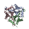
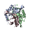
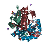
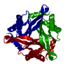
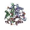
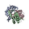
| Title | Crystal structure of stable protein, CutA1, from a psychrotrophic bacterium Shewanella sp. SIB1 | ||||||
 Components Components | CutA1 | ||||||
 Keywords Keywords | ELECTRON TRANSPORT / CutA1 / thermostable protein | ||||||
| Function / homology |  Function and homology information Function and homology information | ||||||
| Biological species |  Shewanella (bacteria) Shewanella (bacteria) | ||||||
| Method |  X-RAY DIFFRACTION / SYNCHROTRON / MOLECULAR REPLACEMENT / Resolution: 2.7 Å X-RAY DIFFRACTION / SYNCHROTRON / MOLECULAR REPLACEMENT / Resolution: 2.7 Å | ||||||
 Authors Authors | Tanaka, S. / Angkawidjaja, C. / Koga, Y. / Takano, K. / Kanaya, S. | ||||||
 Citation Citation | Journal: J.SYNCHROTRON RADIAT. / Year: 2011 Title: Crystal structure of stable protein CutA1 from psychrotrophic bacterium Shewanella sp. SIB1 Authors: Sato, A. / Yokotani, S. / Tadokoro, T. / Tanaka, S.I. / Angkawidjaja, C. / Koga, Y. / Takano, K. / Kanaya, S. | ||||||
| History |
|
- Structure visualization
| Structure viewer | Molecule: MolmilJmol/JSmol |
|---|
- Downloads & links
-Download
| PDBx/mmCIF format | 3ahp.cif.gz | 132.2 KB | Display | PDBx/mmCIF format |
|---|---|---|---|---|
| PDB format | pdb3ahp.ent.gz | 105.6 KB | Display | PDB format |
| PDBx/mmJSON format | 3ahp.json.gz | Tree view | PDBx/mmJSON format | |
| Others |  Other downloads Other downloads |
-Validation report
| Arichive directory | https://data.pdbj.org/pub/pdb/validation_reports/ah/3ahpftp://data.pdbj.org/pub/pdb/validation_reports/ah/3ahp | HTTPS FTP |
|---|
-Related structure data
| Related structure data | 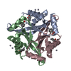 1naqS S: Starting model for refinement |
|---|---|
| Similar structure data |


-Links
 PDBj
PDBj- Assembly
| Deposited unit | 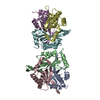
| ||||||||
|---|---|---|---|---|---|---|---|---|---|
| 1 | 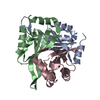
| ||||||||
| 2 | 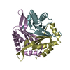
| ||||||||
| Unit cell |
|
-Components
| #1: Protein | Mass: 12098.943 Da / Num. of mol.: 6 Source method: isolated from a genetically manipulated source Source: (gene. exp.) Shewanella (bacteria) / Strain: SIB1 / Plasmid: pET-25b(+) / Production host: #2: Water | ChemComp-HOH / |  Mass: 18.015 Da / Num. of mol.: 63 / Source method: isolated from a natural source / Formula: H2O Mass: 18.015 Da / Num. of mol.: 63 / Source method: isolated from a natural source / Formula: H2OSequence details | A SEQUENCE DATABASE REFERENCE FOR THIS PROTEIN DOES NOT CURRENTLY EXIST. THIS SEQUENCE WILL BE ...A SEQUENCE DATABASE REFERENCE FOR THIS PROTEIN DOES NOT CURRENTLY EXIST. THIS SEQUENCE WILL BE DEPOSITED IN THE SEQUENCE DATABASE. | |
|---|
-Experimental details
-Experiment
| Experiment | Method: X-RAY DIFFRACTION / Number of used crystals: 1 |
|---|
- Sample preparation
| Crystal | Density Matthews: 4.01 Å3/Da / Density % sol: 69.33 % |
|---|---|
| Crystal grow | Temperature: 293 K / Method: vapor diffusion, sitting drop / pH: 4.5 Details: 2.0M Na/K-phosphate, 100mM acetate, pH 4.5, VAPOR DIFFUSION, SITTING DROP, temperature 293K |
-Data collection
| Diffraction | Mean temperature: 100 K |
|---|---|
| Diffraction source | Source: SYNCHROTRON / Site: SPring-8  / Beamline: BL38B1 / Wavelength: 1 Å / Beamline: BL38B1 / Wavelength: 1 Å |
| Detector | Type: ADSC QUANTUM 210 / Detector: CCD / Date: Jun 13, 2009 / Details: mirrors |
| Radiation | Monochromator: Rh mirror / Protocol: SINGLE WAVELENGTH / Monochromatic (M) / Laue (L): M / Scattering type: x-ray |
| Radiation wavelength | Wavelength: 1 Å / Relative weight: 1 |
| Reflection | Resolution: 2.7→50 Å / Num. obs: 33076 / % possible obs: 100 % / Observed criterion σ(F): 0 / Observed criterion σ(I): 0 / Redundancy: 14.6 % / Rmerge(I) obs: 0.175 / Rsym value: 0.143 / Net I/σ(I): 18.8 |
| Reflection shell | Resolution: 2.7→2.75 Å / Redundancy: 14.9 % / Rmerge(I) obs: 0.772 / Mean I/σ(I) obs: 3.03 / Num. unique all: 1627 / Rsym value: 0.689 / % possible all: 100 |
- Processing
| Software |
| |||||||||||||||||||||||||||||||||||||||||||||||||||||||||||||||||
|---|---|---|---|---|---|---|---|---|---|---|---|---|---|---|---|---|---|---|---|---|---|---|---|---|---|---|---|---|---|---|---|---|---|---|---|---|---|---|---|---|---|---|---|---|---|---|---|---|---|---|---|---|---|---|---|---|---|---|---|---|---|---|---|---|---|---|
| Refinement | Method to determine structure: MOLECULAR REPLACEMENT Starting model: PDB ENTRY 1NAQ Resolution: 2.7→30 Å / Cor.coef. Fo:Fc: 0.93 / Cor.coef. Fo:Fc free: 0.901 / SU B: 9.355 / SU ML: 0.199 / Cross valid method: THROUGHOUT / ESU R: 0.381 / ESU R Free: 0.288 Stereochemistry target values: MAXIMUM LIKELIHOOD WITH PHASES Details: HYDROGENS HAVE BEEN ADDED IN THE RIDING POSITIONS
| |||||||||||||||||||||||||||||||||||||||||||||||||||||||||||||||||
| Solvent computation | Ion probe radii: 0.8 Å / Shrinkage radii: 0.8 Å / VDW probe radii: 1.4 Å / Solvent model: MASK | |||||||||||||||||||||||||||||||||||||||||||||||||||||||||||||||||
| Displacement parameters | Biso mean: 35.397 Å2
| |||||||||||||||||||||||||||||||||||||||||||||||||||||||||||||||||
| Refinement step | Cycle: LAST / Resolution: 2.7→30 Å
| |||||||||||||||||||||||||||||||||||||||||||||||||||||||||||||||||
| Refine LS restraints |
| |||||||||||||||||||||||||||||||||||||||||||||||||||||||||||||||||
| LS refinement shell | Resolution: 2.7→2.77 Å / Total num. of bins used: 20
|
