 Movie
Movie Controller

[English] 日本語
 Yorodumi
- PDB-1f1v: ANAEROBIC SUBSTRATE COMPLEX OF HOMOPROTOCATECHUATE 2,3-DIOXYGENAS... -
+ Open data

- Basic information
| Entry | Database: PDB / ID: 1f1v | ||||||
|---|---|---|---|---|---|---|---|
| Title | ANAEROBIC SUBSTRATE COMPLEX OF HOMOPROTOCATECHUATE 2,3-DIOXYGENASE FROM ARTHROBACTER GLOBIFORMIS. (COMPLEX WITH 3,4-DIHYDROXYPHENYLACETATE) | ||||||
 Components Components | HOMOPROTOCATECHUATE 2,3-DIOXYGENASE | ||||||
 Keywords Keywords | OXIDOREDUCTASE / Dioxygenase / Extradiol / Manganese / Biodegradation / Aromatic | ||||||
| Function / homology |  Function and homology information Function and homology information | ||||||
| Biological species |  Arthrobacter globiformis (bacteria) Arthrobacter globiformis (bacteria) | ||||||
| Method |  X-RAY DIFFRACTION / Resolution: 1.9 Å X-RAY DIFFRACTION / Resolution: 1.9 Å | ||||||
 Authors Authors | Vetting, M.W. / Lipscomb, J.D. / Wackett, L.P. / Que Jr., L. / Ohlendorf, D.H. | ||||||
 Citation Citation | Journal: J.Bacteriol. / Year: 2004 Title: Crystallographic comparison of manganese- and iron-dependent homoprotocatechuate 2,3-dioxygenases. Authors: Vetting, M.W. / Wackett, L.P. / Que Jr., L. / Lipscomb, J.D. / Ohlendorf, D.H. #1: Journal: Biochemistry / Year: 1996Title: Manganese(II)-dependent Extradiol-cleaving Catechol Dioxygenase from Arthrobacter globiformis CM-2 Authors: Whiting, A.K. / Boldt, Y.R. / Hendrich, M.P. / Wackett, L.P. / Que Jr, L. #2: Journal: J.BACTERIOL. / Year: 1995Title: A Manganese-dependent Dioxygenase from Arthrobacter globiformis CM-2 belongs to the Major Extradiol Dioxygenase Family Authors: Boldt, Y.R. / Sadowsky, M.J. / Ellis, L.B. / Que Jr, L. / Wackett, L.P. #3: Journal: Biochemistry / Year: 1997Title: Manganese(II) Active Site Mutants of 3,4-dihydroxyphenylacetate 2,3-dioxygenase from Arthrobacter globiformis Strain CM-2. Authors: Boldt, Y.R. / Whiting, A.K. / Wagner, M.L. / Sadowsky, M.J. / Que Jr, L. / Wackett, L.P. #4: Journal: J.Biol.Chem. / Year: 1981Title: 3,4-Dihydroxyphenylacetate 2,3-dioxygenase. A Manganese(II) Dioxygenase from Bacillus brevis. Authors: Que Jr, L. / Widom, J. / Crawford, R.L. | ||||||
| History |
|
- Structure visualization
| Structure viewer | Molecule: MolmilJmol/JSmol |
|---|
- Downloads & links
-Download
| PDBx/mmCIF format | 1f1v.cif.gz | 145.6 KB | Display | PDBx/mmCIF format |
|---|---|---|---|---|
| PDB format | pdb1f1v.ent.gz | 114.2 KB | Display | PDB format |
| PDBx/mmJSON format | 1f1v.json.gz | Tree view | PDBx/mmJSON format | |
| Others |  Other downloads Other downloads |
-Validation report
| Arichive directory | https://data.pdbj.org/pub/pdb/validation_reports/f1/1f1vftp://data.pdbj.org/pub/pdb/validation_reports/f1/1f1v | HTTPS FTP |
|---|
-Related structure data


-Links
 PDBj
PDBj- Assembly
| Deposited unit | 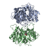
| ||||||||
|---|---|---|---|---|---|---|---|---|---|
| 1 | 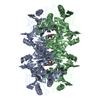
| ||||||||
| Unit cell |
| ||||||||
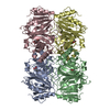
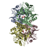
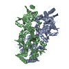
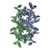
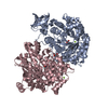
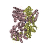
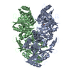
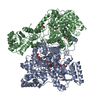
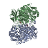
| Details | The biological assembly is a tetramer constructed from chain A and chain B with a symmetry partner generated by the two-fold. |
-Components
| #1: Protein | Mass: 36992.242 Da / Num. of mol.: 2 Source method: isolated from a genetically manipulated source Source: (gene. exp.) Arthrobacter globiformis (bacteria) / Strain: CM-2 / Production host: References: UniProt: Q44048, 3,4-dihydroxyphenylacetate 2,3-dioxygenase #2: Chemical |   Mass: 54.938 Da / Num. of mol.: 2 / Source method: obtained synthetically / Formula: Mn Mass: 54.938 Da / Num. of mol.: 2 / Source method: obtained synthetically / Formula: Mn#3: Chemical | 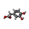  Mass: 168.147 Da / Num. of mol.: 2 / Source method: obtained synthetically / Formula: C8H8O4 / Comment: neurotransmitter*YM Mass: 168.147 Da / Num. of mol.: 2 / Source method: obtained synthetically / Formula: C8H8O4 / Comment: neurotransmitter*YM#4: Water | ChemComp-HOH / |  Mass: 18.015 Da / Num. of mol.: 316 / Source method: isolated from a natural source / Formula: H2O Mass: 18.015 Da / Num. of mol.: 316 / Source method: isolated from a natural source / Formula: H2O |
|---|
-Experimental details
-Experiment
| Experiment | Method: X-RAY DIFFRACTION / Number of used crystals: 1 |
|---|
- Sample preparation
| Crystal | Density Matthews: 2.54 Å3/Da / Density % sol: 51.48 % | ||||||||||||||||||||||||||||||
|---|---|---|---|---|---|---|---|---|---|---|---|---|---|---|---|---|---|---|---|---|---|---|---|---|---|---|---|---|---|---|---|
| Crystal grow | Temperature: 298 K / Method: batch crystallization / pH: 6.8 Details: Peg 8000, Mg Acetate, Na cacodylate, pH 6.8, Batch crystallization, temperature 298K | ||||||||||||||||||||||||||||||
| Crystal grow | *PLUS Temperature: 18 ℃ / pH: 6.5 / Method: batch method | ||||||||||||||||||||||||||||||
| Components of the solutions | *PLUS
|
-Data collection
| Diffraction | Mean temperature: 298 K |
|---|---|
| Diffraction source | Source: ROTATING ANODE / Type: RIGAKU RU200 / Wavelength: 1.5418 |
| Detector | Type: RIGAKU RAXIS IV / Detector: IMAGE PLATE / Date: Jul 2, 1999 |
| Radiation | Protocol: SINGLE WAVELENGTH / Monochromatic (M) / Laue (L): M / Scattering type: x-ray |
| Radiation wavelength | Wavelength: 1.5418 Å / Relative weight: 1 |
| Reflection | Resolution: 1.9→20 Å / Num. all: 58621 / Num. obs: 56746 / % possible obs: 96.8 % / Observed criterion σ(F): 5 / Observed criterion σ(I): 5 / Redundancy: 3.79 % / Rmerge(I) obs: 0.059 / Net I/σ(I): 15.5 |
| Reflection shell | Resolution: 1.9→1.97 Å / Redundancy: 3.8 % / Rmerge(I) obs: 0.063 / Num. unique all: 5438 / % possible all: 94.1 |
| Reflection | *PLUS Highest resolution: 1.9 Å / % possible obs: 96.7 % / Redundancy: 3.8 % |
| Reflection shell | *PLUS % possible obs: 93.6 % / Rmerge(I) obs: 0.236 / Mean I/σ(I) obs: 6.3 |
- Processing
| Software |
| |||||||||||||||||||||||||
|---|---|---|---|---|---|---|---|---|---|---|---|---|---|---|---|---|---|---|---|---|---|---|---|---|---|---|
| Refinement | Resolution: 1.9→20 Å / σ(F): 0 / σ(I): 0 / Stereochemistry target values: Engh & Huber
| |||||||||||||||||||||||||
| Refinement step | Cycle: LAST / Resolution: 1.9→20 Å
| |||||||||||||||||||||||||
| Refine LS restraints |
| |||||||||||||||||||||||||
| Refinement | *PLUS % reflection Rfree: 3 % / Rfactor Rfree: 0.2 | |||||||||||||||||||||||||
| Solvent computation | *PLUS | |||||||||||||||||||||||||
| Displacement parameters | *PLUS | |||||||||||||||||||||||||
| Refine LS restraints | *PLUS
| |||||||||||||||||||||||||
| LS refinement shell | *PLUS Rfactor Rfree: 0.296 / Rfactor Rwork: 0.252 |
