 Movie
Movie Controller

[English] 日本語
 Yorodumi
- PDB-1f1x: CRYSTAL STRUCTURE OF HOMOPROTOCATECHUATE 2,3-DIOXYGENASE FROM BRE... -
+ Open data

- Basic information
| Entry | Database: PDB / ID: 1f1x | ||||||
|---|---|---|---|---|---|---|---|
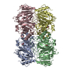
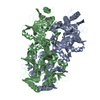
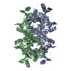
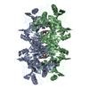
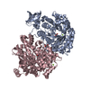
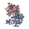
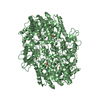
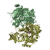
| Title | CRYSTAL STRUCTURE OF HOMOPROTOCATECHUATE 2,3-DIOXYGENASE FROM BREVIBACTERIUM FUSCUM | ||||||
 Components Components | HOMOPROTOCATECHUATE 2,3-DIOXYGENASE | ||||||
 Keywords Keywords | OXIDOREDUCTASE / Dioxygenase / Extradiol / Iron / Biodegradation / Aromatic | ||||||
| Function / homology |  Function and homology information Function and homology information | ||||||
| Biological species |  Brevibacterium fuscum (bacteria) Brevibacterium fuscum (bacteria) | ||||||
| Method |  X-RAY DIFFRACTION / SYNCHROTRON / Resolution: 1.6 Å X-RAY DIFFRACTION / SYNCHROTRON / Resolution: 1.6 Å | ||||||
 Authors Authors | Vetting, M.W. / Lipscomb, J.D. / Wackett, L.P. / Que Jr., L. / Ohlendorf, D.H. | ||||||
 Citation Citation | Journal: J.Bacteriol. / Year: 2004 Title: Crystallographic comparison of manganese- and iron-dependent homoprotocatechuate 2,3-dioxygenases. Authors: Vetting, M.W. / Wackett, L.P. / Que Jr., L. / Lipscomb, J.D. / Ohlendorf, D.H. #1: Journal: J.Biol.Chem. / Year: 1996Title: Homoprotocatechuate 2,3-dioxygenase from Brevibacterium fuscum. A Dioxygenase with Catalase Activity. Authors: Miller, M.A. / Lipscomb, J.D. #2: Journal: Protein Expr.Purif. / Year: 1997Title: Cloning, overexpression, and mutagenesis of the gene for homoprotocatechuate 2,3-dioxygenase from Brevibacterium fuscum. Authors: Wang, Y.Z. / Lipscomb, J.D. | ||||||
| History |
|
- Structure visualization
| Structure viewer | Molecule: MolmilJmol/JSmol |
|---|
- Downloads & links
-Download
| PDBx/mmCIF format | 1f1x.cif.gz | 285.8 KB | Display | PDBx/mmCIF format |
|---|---|---|---|---|
| PDB format | pdb1f1x.ent.gz | 230 KB | Display | PDB format |
| PDBx/mmJSON format | 1f1x.json.gz | Tree view | PDBx/mmJSON format | |
| Others |  Other downloads Other downloads |
-Validation report
| Arichive directory | https://data.pdbj.org/pub/pdb/validation_reports/f1/1f1xftp://data.pdbj.org/pub/pdb/validation_reports/f1/1f1x | HTTPS FTP |
|---|
-Related structure data


-Links
 PDBj
PDBj- Assembly
| Deposited unit | 
| ||||||||
|---|---|---|---|---|---|---|---|---|---|
| 1 |
| ||||||||
| Unit cell |
| ||||||||
| Details | The biological assembly is a tetramer. There is a tetramer per asymmetric unit. |
-Components
| #1: Protein | Mass: 37161.516 Da / Num. of mol.: 4 Source method: isolated from a genetically manipulated source Source: (gene. exp.) Brevibacterium fuscum (bacteria) / Production host: References: UniProt: Q45135, 3,4-dihydroxyphenylacetate 2,3-dioxygenase #2: Chemical | ChemComp-FEL /   Mass: 109.891 Da / Num. of mol.: 4 / Source method: obtained synthetically / Formula: FeH6O3 Mass: 109.891 Da / Num. of mol.: 4 / Source method: obtained synthetically / Formula: FeH6O3#3: Water | ChemComp-HOH / |  Mass: 18.015 Da / Num. of mol.: 1093 / Source method: isolated from a natural source / Formula: H2O Mass: 18.015 Da / Num. of mol.: 1093 / Source method: isolated from a natural source / Formula: H2O |
|---|
-Experimental details
-Experiment
| Experiment | Method: X-RAY DIFFRACTION / Number of used crystals: 1 |
|---|
- Sample preparation
| Crystal | Density Matthews: 2.52 Å3/Da / Density % sol: 51.12 % | ||||||||||||||||||||||||||||||
|---|---|---|---|---|---|---|---|---|---|---|---|---|---|---|---|---|---|---|---|---|---|---|---|---|---|---|---|---|---|---|---|
| Crystal grow | Temperature: 298 K / Method: batch crystallization / pH: 7.5 Details: Peg5000, Mg Acetate, MOPS, Glycerol was add prior to data collection., pH 7.5, Batch crystallization, temperature 298K | ||||||||||||||||||||||||||||||
| Crystal grow | *PLUS Temperature: 18 ℃ / Method: vapor diffusion, hanging drop | ||||||||||||||||||||||||||||||
| Components of the solutions | *PLUS
|
-Data collection
| Diffraction | Mean temperature: 200 K |
|---|---|
| Diffraction source | Source: SYNCHROTRON / Site: APS  / Beamline: 19-ID / Wavelength: 1.032 / Beamline: 19-ID / Wavelength: 1.032 |
| Detector | Type: CUSTOM-MADE / Detector: CCD / Date: Feb 7, 1998 |
| Radiation | Protocol: SINGLE WAVELENGTH / Monochromatic (M) / Laue (L): M / Scattering type: x-ray |
| Radiation wavelength | Wavelength: 1.032 Å / Relative weight: 1 |
| Reflection | Resolution: 1.5→30 Å / Num. all: 234020 / Num. obs: 216266 / % possible obs: 92.4 % / Observed criterion σ(F): 5 / Observed criterion σ(I): 5 / Redundancy: 2.54 % / Rmerge(I) obs: 0.052 / Net I/σ(I): 20 |
| Reflection shell | Resolution: 1.5→1.55 Å / Redundancy: 1.5 % / Rmerge(I) obs: 0.094 / Num. unique all: 19027 / % possible all: 81.2 |
| Reflection | *PLUS Lowest resolution: 20 Å / % possible obs: 92.9 % / Redundancy: 2.55 % / Rmerge(I) obs: 0.041 |
| Reflection shell | *PLUS % possible obs: 79.5 % / Redundancy: 1.51 % / Rmerge(I) obs: 0.127 / Mean I/σ(I) obs: 8.7 |
- Processing
| Software |
| |||||||||||||||||||||||||
|---|---|---|---|---|---|---|---|---|---|---|---|---|---|---|---|---|---|---|---|---|---|---|---|---|---|---|
| Refinement | Resolution: 1.6→30 Å / σ(F): 0 / σ(I): 0 / Stereochemistry target values: Engh & Huber
| |||||||||||||||||||||||||
| Refinement step | Cycle: LAST / Resolution: 1.6→30 Å
| |||||||||||||||||||||||||
| Refine LS restraints |
| |||||||||||||||||||||||||
| Refinement | *PLUS Lowest resolution: 20 Å / % reflection Rfree: 10 % | |||||||||||||||||||||||||
| Solvent computation | *PLUS | |||||||||||||||||||||||||
| Displacement parameters | *PLUS | |||||||||||||||||||||||||
| Refine LS restraints | *PLUS
| |||||||||||||||||||||||||
| LS refinement shell | *PLUS Rfactor Rfree: 0.239 / Rfactor Rwork: 0.187 |
