 Movie
Movie Controller

[English] 日本語
 Yorodumi
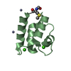
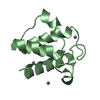
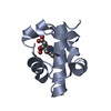
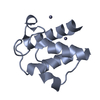
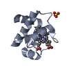
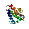
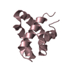
- PDB-1x3o: Crystal structure of the acyl carrier protein from thermus thermo... -
+ Open data

- Basic information
| Entry | Database: PDB / ID: 1x3o | ||||||
|---|---|---|---|---|---|---|---|
| Title | Crystal structure of the acyl carrier protein from thermus thermophilus HB8 | ||||||
 Components Components | acyl carrier protein | ||||||
 Keywords Keywords | LIPID TRANSPORT / structural genomics / RIKEN Structural Genomics/Proteomics Initiative / RSGI / NPPSFA / National Project on Protein Structural and Functional Analyses | ||||||
| Function / homology |  Function and homology information Function and homology informationlipid A biosynthetic process / acyl binding / acyl carrier activity / membrane / cytosol Similarity search - Function | ||||||
| Biological species |   Thermus thermophilus (bacteria) Thermus thermophilus (bacteria) | ||||||
| Method |  X-RAY DIFFRACTION / SYNCHROTRON / MOLECULAR REPLACEMENT / Resolution: 1.5 Å X-RAY DIFFRACTION / SYNCHROTRON / MOLECULAR REPLACEMENT / Resolution: 1.5 Å | ||||||
 Authors Authors | Mizutani, H. / Kunishima, N. / RIKEN Structural Genomics/Proteomics Initiative (RSGI) | ||||||
 Citation Citation | Journal: To be Published Title: Crystal structure of the acyl carrier protein from thermus thermophilus HB8 Authors: Mizutani, H. / Kunishima, N. | ||||||
| History |
|
- Structure visualization
| Structure viewer | Molecule: MolmilJmol/JSmol |
|---|
- Downloads & links
-Download
| PDBx/mmCIF format | 1x3o.cif.gz | 28.7 KB | Display | PDBx/mmCIF format |
|---|---|---|---|---|
| PDB format | pdb1x3o.ent.gz | 18.9 KB | Display | PDB format |
| PDBx/mmJSON format | 1x3o.json.gz | Tree view | PDBx/mmJSON format | |
| Others |  Other downloads Other downloads |
-Validation report
| Summary document | 1x3o_validation.pdf.gz | 422 KB | Display | wwPDB validaton report |
|---|---|---|---|---|
| Full document | 1x3o_full_validation.pdf.gz | 423.6 KB | Display | |
| Data in XML | 1x3o_validation.xml.gz | 6.5 KB | Display | |
| Data in CIF | 1x3o_validation.cif.gz | 8.3 KB | Display | |
| Arichive directory | https://data.pdbj.org/pub/pdb/validation_reports/x3/1x3oftp://data.pdbj.org/pub/pdb/validation_reports/x3/1x3o | HTTPS FTP |
-Related structure data
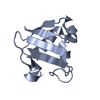
| Related structure data |  1l0hS S: Starting model for refinement |
|---|---|
| Similar structure data | |
| Other databases |

-Links
 PDBj
PDBj
- Assembly
| Deposited unit | 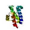
| ||||||||
|---|---|---|---|---|---|---|---|---|---|
| 1 |
| ||||||||
| Unit cell |
| ||||||||
| Components on special symmetry positions |
| ||||||||
| Details | The biological assembly is monomer. |
-Components
| #1: Protein | Mass: 9024.226 Da / Num. of mol.: 1 Source method: isolated from a genetically manipulated source Source: (gene. exp.) Thermus thermophilus (bacteria) / Strain: HB8 / Plasmid: pET11a / Species (production host): Escherichia coli / Production host: |
|---|---|
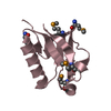
| #2: Water | ChemComp-HOH /  Mass: 18.015 Da / Num. of mol.: 100 / Source method: isolated from a natural source / Formula: H2O Mass: 18.015 Da / Num. of mol.: 100 / Source method: isolated from a natural source / Formula: H2O |
-Experimental details
-Experiment
| Experiment | Method: X-RAY DIFFRACTION / Number of used crystals: 1 |
|---|
- Sample preparation
| Crystal | Density Matthews: 3.15 Å3/Da / Density % sol: 60.94 % |
|---|---|
| Crystal grow | Temperature: 293 K / Method: gel-tube / pH: 4.2 Details: PEG 4000, dioxane, acetate, pH 4.2, Gel-Tube, temperature 293K |
-Data collection
| Diffraction | Mean temperature: 100 K |
|---|---|
| Diffraction source | Source: SYNCHROTRON / Site: SPring-8  / Beamline: BL26B1 / Wavelength: 0.9 Å / Beamline: BL26B1 / Wavelength: 0.9 Å |
| Detector | Type: RIGAKU JUPITER 210 / Detector: CCD / Date: Nov 11, 2004 / Details: mirrors |
| Radiation | Monochromator: Bending magnet / Protocol: SINGLE WAVELENGTH / Monochromatic (M) / Laue (L): M / Scattering type: x-ray |
| Radiation wavelength | Wavelength: 0.9 Å / Relative weight: 1 |
| Reflection | Resolution: 1.5→30 Å / Num. all: 18911 / Num. obs: 18911 / % possible obs: 98.8 % / Observed criterion σ(F): 0 / Observed criterion σ(I): 0 / Redundancy: 10.1 % / Biso Wilson estimate: 24.4 Å2 / Rmerge(I) obs: 0.046 / Rsym value: 0.045 / Net I/σ(I): 16.7 |
| Reflection shell | Resolution: 1.5→1.55 Å / Redundancy: 10.2 % / Rmerge(I) obs: 0.473 / Mean I/σ(I) obs: 4.06 / Num. unique all: 1799 / Rsym value: 0.453 / % possible all: 96 |
- Processing
| Software |
| ||||||||||||||||||||||||||||||||||||||||||||||||||||||||||||||||||||||||||||||||
|---|---|---|---|---|---|---|---|---|---|---|---|---|---|---|---|---|---|---|---|---|---|---|---|---|---|---|---|---|---|---|---|---|---|---|---|---|---|---|---|---|---|---|---|---|---|---|---|---|---|---|---|---|---|---|---|---|---|---|---|---|---|---|---|---|---|---|---|---|---|---|---|---|---|---|---|---|---|---|---|---|---|
| Refinement | Method to determine structure: MOLECULAR REPLACEMENT Starting model: PDB ENTRY 1L0H Resolution: 1.5→30 Å / Rfactor Rfree error: 0.009 / Data cutoff high absF: 909289.74 / Data cutoff low absF: 0 / Isotropic thermal model: RESTRAINED / Cross valid method: THROUGHOUT / σ(F): 0 / Stereochemistry target values: Engh & Huber
| ||||||||||||||||||||||||||||||||||||||||||||||||||||||||||||||||||||||||||||||||
| Solvent computation | Solvent model: FLAT MODEL / Bsol: 40.1365 Å2 / ksol: 0.353233 e/Å3 | ||||||||||||||||||||||||||||||||||||||||||||||||||||||||||||||||||||||||||||||||
| Displacement parameters | Biso mean: 26.2 Å2
| ||||||||||||||||||||||||||||||||||||||||||||||||||||||||||||||||||||||||||||||||
| Refine analyze |
| ||||||||||||||||||||||||||||||||||||||||||||||||||||||||||||||||||||||||||||||||
| Refinement step | Cycle: LAST / Resolution: 1.5→30 Å
| ||||||||||||||||||||||||||||||||||||||||||||||||||||||||||||||||||||||||||||||||
| Refine LS restraints |
| ||||||||||||||||||||||||||||||||||||||||||||||||||||||||||||||||||||||||||||||||
| LS refinement shell | Resolution: 1.5→1.59 Å / Rfactor Rfree error: 0.029 / Total num. of bins used: 6
| ||||||||||||||||||||||||||||||||||||||||||||||||||||||||||||||||||||||||||||||||
| Xplor file |
|
