 Movie
Movie Controller

[English] 日本語
 Yorodumi
- PDB-1x25: Crystal Structure of a Member of YjgF Family from Sulfolobus Toko... -
+ Open data

- Basic information
| Entry | Database: PDB / ID: 1x25 | ||||||
|---|---|---|---|---|---|---|---|
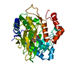
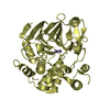
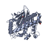
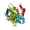
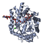
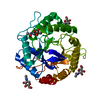
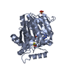
| Title | Crystal Structure of a Member of YjgF Family from Sulfolobus Tokodaii (ST0811) | ||||||
 Components Components | Hypothetical UPF0076 protein ST0811 | ||||||
 Keywords Keywords | STRUCTURAL GENOMICS / UNKNOWN FUNCTION / YJGF-LIKE PROTEIN / ARCHAEA / ST0811 | ||||||
| Function / homology |  Function and homology information Function and homology information | ||||||
| Biological species |   Sulfolobus tokodaii (archaea) Sulfolobus tokodaii (archaea) | ||||||
| Method |  X-RAY DIFFRACTION / MOLECULAR REPLACEMENT / Resolution: 2 Å X-RAY DIFFRACTION / MOLECULAR REPLACEMENT / Resolution: 2 Å | ||||||
 Authors Authors | Miyakawa, T. / Lee, W.C. / Hatano, K. / Kato, Y. / Sawano, Y. / Miyazono, K. / Nagata, K. / Tanokura, M. | ||||||
 Citation Citation | Journal: Proteins / Year: 2006 Title: Crystal structure of the YjgF/YER057c/UK114 family protein from the hyperthermophilic archaeon Sulfolobus tokodaii strain 7 Authors: Miyakawa, T. / Lee, W.C. / Hatano, K. / Kato, Y. / Sawano, Y. / Miyazono, K. / Nagata, K. / Tanokura, M. | ||||||
| History |
|
- Structure visualization
| Structure viewer | Molecule: MolmilJmol/JSmol |
|---|
- Downloads & links
-Download
| PDBx/mmCIF format | 1x25.cif.gz | 65.4 KB | Display | PDBx/mmCIF format |
|---|---|---|---|---|
| PDB format | pdb1x25.ent.gz | 48.6 KB | Display | PDB format |
| PDBx/mmJSON format | 1x25.json.gz | Tree view | PDBx/mmJSON format | |
| Others |  Other downloads Other downloads |
-Validation report
| Arichive directory | https://data.pdbj.org/pub/pdb/validation_reports/x2/1x25ftp://data.pdbj.org/pub/pdb/validation_reports/x2/1x25 | HTTPS FTP |
|---|
-Related structure data
| Related structure data |  1qd9S S: Starting model for refinement |
|---|---|
| Similar structure data |


-Links
 PDBj
PDBj
- Assembly
| Deposited unit | 
| ||||||||||||
|---|---|---|---|---|---|---|---|---|---|---|---|---|---|
| 1 | 
| ||||||||||||
| 2 | 
| ||||||||||||
| 3 | 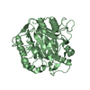
| ||||||||||||
| Unit cell |
| ||||||||||||
| Components on special symmetry positions |
| ||||||||||||
| Details | The biological assembly is a trimer generated from a molecule in the asymmetric unit by the operations: -y, x-y, z and -x+y, -x, z. |
-Components
| #1: Protein | Mass: 14155.356 Da / Num. of mol.: 2 Source method: isolated from a genetically manipulated source Source: (gene. exp.) Sulfolobus tokodaii (archaea) / Strain: str. 7 / Gene: ST0811 / Plasmid: pET28a / Production host:  #2: Water | ChemComp-HOH / |  Mass: 18.015 Da / Num. of mol.: 210 / Source method: isolated from a natural source / Formula: H2O Mass: 18.015 Da / Num. of mol.: 210 / Source method: isolated from a natural source / Formula: H2O |
|---|
-Experimental details
-Experiment
| Experiment | Method: X-RAY DIFFRACTION / Number of used crystals: 1 |
|---|
- Sample preparation
| Crystal | Density Matthews: 2.33 Å3/Da / Density % sol: 46.7 % |
|---|---|
| Crystal grow | Temperature: 293 K / Method: vapor diffusion, sitting drop / pH: 5.3 Details: PEG 10000, Bis-Tris, ammonium acetate, pH 5.3, VAPOR DIFFUSION, SITTING DROP, temperature 293K |
-Data collection
| Diffraction | Mean temperature: 100 K |
|---|---|
| Diffraction source | Source: ROTATING ANODE / Type: RIGAKU FR-E / Wavelength: 1.5418 Å |
| Detector | Type: RIGAKU RAXIS VII / Detector: IMAGE PLATE / Date: Sep 1, 2004 |
| Radiation | Monochromator: CONFOCAL MIRROR / Protocol: SINGLE WAVELENGTH / Monochromatic (M) / Laue (L): M / Scattering type: x-ray |
| Radiation wavelength | Wavelength: 1.5418 Å / Relative weight: 1 |
| Reflection | Resolution: 2→22.051 Å / Num. all: 16882 / Num. obs: 16882 / % possible obs: 99.8 % / Observed criterion σ(I): 5.1 / Redundancy: 6.2 % / Biso Wilson estimate: 17.336 Å2 / Rmerge(I) obs: 0.061 / Rsym value: 0.055 / Net I/σ(I): 9.8 |
| Reflection shell | Resolution: 2→2.11 Å / Redundancy: 5.9 % / Rmerge(I) obs: 0.151 / Mean I/σ(I) obs: 5.1 / Num. unique all: 182 / Rsym value: 0.137 / % possible all: 99.8 |
- Processing
| Software |
| ||||||||||||||||||||||||||||||||||||||||||||||||||||||||||||||||||||||
|---|---|---|---|---|---|---|---|---|---|---|---|---|---|---|---|---|---|---|---|---|---|---|---|---|---|---|---|---|---|---|---|---|---|---|---|---|---|---|---|---|---|---|---|---|---|---|---|---|---|---|---|---|---|---|---|---|---|---|---|---|---|---|---|---|---|---|---|---|---|---|---|
| Refinement | Method to determine structure: MOLECULAR REPLACEMENT Starting model: PDB ENTRY 1QD9 Resolution: 2→19.03 Å / Cor.coef. Fo:Fc: 0.96 / Cor.coef. Fo:Fc free: 0.94 / SU B: 2.786 / SU ML: 0.082 / Cross valid method: THROUGHOUT / ESU R: 0.163 / ESU R Free: 0.141 / Stereochemistry target values: MAXIMUM LIKELIHOOD
| ||||||||||||||||||||||||||||||||||||||||||||||||||||||||||||||||||||||
| Solvent computation | Ion probe radii: 0.8 Å / Shrinkage radii: 0.8 Å / VDW probe radii: 1.4 Å / Solvent model: BABINET MODEL WITH MASK | ||||||||||||||||||||||||||||||||||||||||||||||||||||||||||||||||||||||
| Displacement parameters | Biso mean: 18.36 Å2
| ||||||||||||||||||||||||||||||||||||||||||||||||||||||||||||||||||||||
| Refinement step | Cycle: LAST / Resolution: 2→19.03 Å
| ||||||||||||||||||||||||||||||||||||||||||||||||||||||||||||||||||||||
| Refine LS restraints |
| ||||||||||||||||||||||||||||||||||||||||||||||||||||||||||||||||||||||
| LS refinement shell | Resolution: 2→2.052 Å / Total num. of bins used: 20
|
