 Movie
Movie Controller

[English] 日本語
 Yorodumi
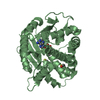
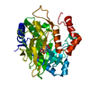
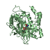
- PDB-5lun: Ethylene Forming Enzyme from Pseudomonas syringae pv. phaseolicol... -
+ Open data

- Basic information
| Entry | Database: PDB / ID: 5lun | ||||||
|---|---|---|---|---|---|---|---|
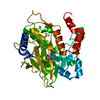
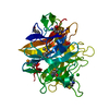
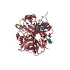
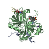
| Title | Ethylene Forming Enzyme from Pseudomonas syringae pv. phaseolicola - P1 ultra-high resolution crystal form in complex with iron, N-oxalylglycine and arginine | ||||||
 Components Components | 2-oxoglutarate-dependent ethylene/succinate-forming enzyme | ||||||
 Keywords Keywords | OXIDOREDUCTASE / 2-oxoglutarate and ferrous iron dependent oxygenase / ethylene forming / double stranded beta helix | ||||||
| Function / homology |  Function and homology information Function and homology information2-oxoglutarate dioxygenase (ethene-forming) / 2-oxoglutarate/L-arginine monooxygenase/decarboxylase (succinate-forming) / 2-oxoglutarate oxygenase/decarboxylase (ethylene-forming) activity / ethylene biosynthetic process / dioxygenase activity / metal ion binding Similarity search - Function | ||||||
| Biological species |  Pseudomonas savastanoi pv. phaseolicola (bacteria) Pseudomonas savastanoi pv. phaseolicola (bacteria) | ||||||
| Method |  X-RAY DIFFRACTION / SYNCHROTRON / MOLECULAR REPLACEMENT / molecular replacement / Resolution: 1.08 Å X-RAY DIFFRACTION / SYNCHROTRON / MOLECULAR REPLACEMENT / molecular replacement / Resolution: 1.08 Å | ||||||
 Authors Authors | McDonough, M.A. / Zhang, Z. / Schofield, C.J. | ||||||
 Citation Citation | Journal: Proc. Natl. Acad. Sci. U.S.A. / Year: 2017 Title: Structural and stereoelectronic insights into oxygenase-catalyzed formation of ethylene from 2-oxoglutarate. Authors: Zhang, Z. / Smart, T.J. / Choi, H. / Hardy, F. / Lohans, C.T. / Abboud, M.I. / Richardson, M.S.W. / Paton, R.S. / McDonough, M.A. / Schofield, C.J. | ||||||
| History |
|
- Structure visualization
| Structure viewer | Molecule: MolmilJmol/JSmol |
|---|
- Downloads & links
-Download
| PDBx/mmCIF format | 5lun.cif.gz | 607.5 KB | Display | PDBx/mmCIF format |
|---|---|---|---|---|
| PDB format | pdb5lun.ent.gz | 491.4 KB | Display | PDB format |
| PDBx/mmJSON format | 5lun.json.gz | Tree view | PDBx/mmJSON format | |
| Others |  Other downloads Other downloads |
-Validation report
| Arichive directory | https://data.pdbj.org/pub/pdb/validation_reports/lu/5lunftp://data.pdbj.org/pub/pdb/validation_reports/lu/5lun | HTTPS FTP |
|---|
-Related structure data
| Related structure data |  5lsqSC  5mofC S: Starting model for refinement C: citing same article ( |
|---|---|
| Similar structure data |


-Links
 PDBj
PDBj

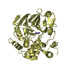
- Assembly
| Deposited unit | 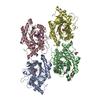
| ||||||||
|---|---|---|---|---|---|---|---|---|---|
| 1 | 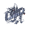
| ||||||||
| 2 | 
| ||||||||
| 3 | 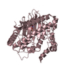
| ||||||||
| 4 | 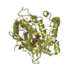
| ||||||||
| Unit cell |
|
-Components
-Protein , 1 types, 4 molecules ABCD
| #1: Protein | Mass: 39632.602 Da / Num. of mol.: 4 Source method: isolated from a genetically manipulated source Source: (gene. exp.) Pseudomonas savastanoi pv. phaseolicola (bacteria)Gene: efe / Plasmid: pColdI / Production host: References: UniProt: P32021, 2-oxoglutarate dioxygenase (ethene-forming), EC: 1.14.11.34 |
|---|
-Non-polymers , 5 types, 2065 molecules 


| #2: Chemical | ChemComp-FE /  Mass: 55.845 Da / Num. of mol.: 4 / Source method: obtained synthetically / Formula: Fe Mass: 55.845 Da / Num. of mol.: 4 / Source method: obtained synthetically / Formula: Fe#3: Chemical | ChemComp-OGA /  Mass: 147.086 Da / Num. of mol.: 4 / Source method: obtained synthetically / Formula: C4H5NO5 / Comment: inhibitor*YM Mass: 147.086 Da / Num. of mol.: 4 / Source method: obtained synthetically / Formula: C4H5NO5 / Comment: inhibitor*YM#4: Chemical | ChemComp-ARG /  Type: L-peptide linking / Mass: 175.209 Da / Num. of mol.: 4 / Source method: obtained synthetically / Formula: C6H15N4O2 Type: L-peptide linking / Mass: 175.209 Da / Num. of mol.: 4 / Source method: obtained synthetically / Formula: C6H15N4O2#5: Chemical |  Mass: 92.094 Da / Num. of mol.: 3 / Source method: obtained synthetically / Formula: C3H8O3 Mass: 92.094 Da / Num. of mol.: 3 / Source method: obtained synthetically / Formula: C3H8O3#6: Water | ChemComp-HOH / | Mass: 18.015 Da / Num. of mol.: 2050 / Source method: isolated from a natural source / Formula: H2O |
|---|
-Experimental details
-Experiment
| Experiment | Method: X-RAY DIFFRACTION / Number of used crystals: 1 |
|---|
- Sample preparation
| Crystal | Density Matthews: 2.38 Å3/Da / Density % sol: 48.38 % |
|---|---|
| Crystal grow | Temperature: 298 K / Method: vapor diffusion, hanging drop / pH: 8 Details: 20% polyethylene glycol, 0.1 M Tris, 0.2 M sodium chloride, 0.01 M N-oxalylglycine, 0.02 M L-Arginine, protein concentration 15mg/mL |
-Data collection
| Diffraction | Mean temperature: 100 K |
|---|---|
| Diffraction source | Source: SYNCHROTRON / Site: Diamond  / Beamline: I02 / Wavelength: 0.97949 Å / Beamline: I02 / Wavelength: 0.97949 Å |
| Detector | Type: DECTRIS PILATUS 6M-F / Detector: PIXEL / Date: May 25, 2015 |
| Radiation | Protocol: SINGLE WAVELENGTH / Monochromatic (M) / Laue (L): M / Scattering type: x-ray |
| Radiation wavelength | Wavelength: 0.97949 Å / Relative weight: 1 |
| Reflection | Resolution: 1.08→77.6 Å / Num. obs: 584165 / % possible obs: 93 % / Redundancy: 3.5 % / Biso Wilson estimate: 11.557 Å2 / CC1/2: 0.994 / Rmerge(I) obs: 0.053 / Net I/σ(I): 7.7 |
| Reflection shell | Resolution: 1.08→1.12 Å / Redundancy: 3.5 % / Rmerge(I) obs: 0.822 / Mean I/σ(I) obs: 1.4 / CC1/2: 0.701 / % possible all: 89.3 |
-Phasing
| Phasing | Method: molecular replacement |
|---|
- Processing
| Software |
| |||||||||||||||||||||||||||||||||||||||||||||||||||||||||||||||||||||||||||||||||||||||||||||||||||||||||||||||||||||||||||||||||||||||||||||||||||||||||||||||||||||||||||||||||||||||||||||||||||||||||||||||||||||||||
|---|---|---|---|---|---|---|---|---|---|---|---|---|---|---|---|---|---|---|---|---|---|---|---|---|---|---|---|---|---|---|---|---|---|---|---|---|---|---|---|---|---|---|---|---|---|---|---|---|---|---|---|---|---|---|---|---|---|---|---|---|---|---|---|---|---|---|---|---|---|---|---|---|---|---|---|---|---|---|---|---|---|---|---|---|---|---|---|---|---|---|---|---|---|---|---|---|---|---|---|---|---|---|---|---|---|---|---|---|---|---|---|---|---|---|---|---|---|---|---|---|---|---|---|---|---|---|---|---|---|---|---|---|---|---|---|---|---|---|---|---|---|---|---|---|---|---|---|---|---|---|---|---|---|---|---|---|---|---|---|---|---|---|---|---|---|---|---|---|---|---|---|---|---|---|---|---|---|---|---|---|---|---|---|---|---|---|---|---|---|---|---|---|---|---|---|---|---|---|---|---|---|---|---|---|---|---|---|---|---|---|---|---|---|---|---|---|---|---|
| Refinement | Method to determine structure: MOLECULAR REPLACEMENT Starting model: 5LSQ Resolution: 1.08→77.597 Å / SU ML: 0.15 / Cross valid method: THROUGHOUT / σ(F): 1.96 / Phase error: 26.14
| |||||||||||||||||||||||||||||||||||||||||||||||||||||||||||||||||||||||||||||||||||||||||||||||||||||||||||||||||||||||||||||||||||||||||||||||||||||||||||||||||||||||||||||||||||||||||||||||||||||||||||||||||||||||||
| Solvent computation | Shrinkage radii: 0.9 Å / VDW probe radii: 1.11 Å | |||||||||||||||||||||||||||||||||||||||||||||||||||||||||||||||||||||||||||||||||||||||||||||||||||||||||||||||||||||||||||||||||||||||||||||||||||||||||||||||||||||||||||||||||||||||||||||||||||||||||||||||||||||||||
| Displacement parameters | Biso max: 75.14 Å2 / Biso mean: 21.1423 Å2 / Biso min: 6.3 Å2 | |||||||||||||||||||||||||||||||||||||||||||||||||||||||||||||||||||||||||||||||||||||||||||||||||||||||||||||||||||||||||||||||||||||||||||||||||||||||||||||||||||||||||||||||||||||||||||||||||||||||||||||||||||||||||
| Refinement step | Cycle: final / Resolution: 1.08→77.597 Å
| |||||||||||||||||||||||||||||||||||||||||||||||||||||||||||||||||||||||||||||||||||||||||||||||||||||||||||||||||||||||||||||||||||||||||||||||||||||||||||||||||||||||||||||||||||||||||||||||||||||||||||||||||||||||||
| Refine LS restraints |
| |||||||||||||||||||||||||||||||||||||||||||||||||||||||||||||||||||||||||||||||||||||||||||||||||||||||||||||||||||||||||||||||||||||||||||||||||||||||||||||||||||||||||||||||||||||||||||||||||||||||||||||||||||||||||
| LS refinement shell | Refine-ID: X-RAY DIFFRACTION / Total num. of bins used: 30
|
