 ムービー
ムービー コントローラー


+ データを開く

- 基本情報
基本情報
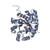
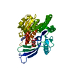
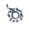
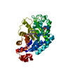
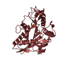
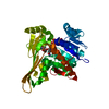
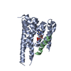
| 登録情報 | データベース: PDB / ID: 1wq5 | ||||||
|---|---|---|---|---|---|---|---|
| タイトル | Crystal structure of tryptophan synthase alpha-subunit from Escherichia coli | ||||||
 要素 要素 | Tryptophan synthase alpha chain | ||||||
 キーワード キーワード | LYASE / Tryptophan synthase / tryptophan / RIKEN Structural Genomics/Proteomics Initiative / RSGI / Structural Genomics | ||||||
| 機能・相同性 |  機能・相同性情報 機能・相同性情報tryptophan synthase / tryptophan synthase activity / L-tryptophan biosynthetic process / aromatic amino acid family biosynthetic process / molecular adaptor activity / lyase activity / cytoplasm / cytosol 類似検索 - 分子機能 | ||||||
| 生物種 |  | ||||||
| 手法 |  X線回折 / シンクロトロン / 分子置換 / 解像度: 2.3 Å X線回折 / シンクロトロン / 分子置換 / 解像度: 2.3 Å | ||||||
 データ登録者 データ登録者 | Nishio, K. / Morimoto, Y. / Ishizuka, M. / Ogasahara, K. / Yutani, K. / Tsukihara, T. / RIKEN Structural Genomics/Proteomics Initiative (RSGI) | ||||||
 引用 引用 | ジャーナル: Biochemistry / 年: 2005 タイトル: Conformational Changes in the alpha-Subunit Coupled to Binding of the beta(2)-Subunit of Tryptophan Synthase from Escherichia coli: Crystal Structure of the Tryptophan Synthase alpha-Subunit Alon 著者: Nishio, K. / Morimoto, Y. / Ishizuka, M. / Ogasahara, K. / Tsukihara, T. / Yutani, K. | ||||||
| 履歴 |
|
- 構造の表示
構造の表示
| 構造ビューア | 分子: MolmilJmol/JSmol |
|---|
- ダウンロードとリンク
-ダウンロード
| PDBx/mmCIF形式 | 1wq5.cif.gz | 117.6 KB | 表示 | PDBx/mmCIF形式 |
|---|---|---|---|---|
| PDB形式 | pdb1wq5.ent.gz | 91.9 KB | 表示 | PDB形式 |
| PDBx/mmJSON形式 | 1wq5.json.gz | ツリー表示 | PDBx/mmJSON形式 | |
| その他 |  その他のダウンロード その他のダウンロード |
-検証レポート
| 文書・要旨 | 1wq5_validation.pdf.gz | 462.3 KB | 表示 | wwPDB検証レポート |
|---|---|---|---|---|
| 文書・詳細版 | 1wq5_full_validation.pdf.gz | 472.8 KB | 表示 | |
| XML形式データ | 1wq5_validation.xml.gz | 23.8 KB | 表示 | |
| CIF形式データ | 1wq5_validation.cif.gz | 32.9 KB | 表示 | |
| アーカイブディレクトリ | https://data.pdbj.org/pub/pdb/validation_reports/wq/1wq5ftp://data.pdbj.org/pub/pdb/validation_reports/wq/1wq5 | HTTPS FTP |
-関連構造データ


-リンク
 PDBj
PDBj
- 集合体
集合体
| 登録構造単位 | 
| ||||||||
|---|---|---|---|---|---|---|---|---|---|
| 1 | 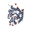
| ||||||||
| 2 | 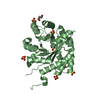
| ||||||||
| 3 | 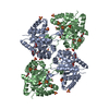
| ||||||||
| 単位格子 |
|
-要素
| #1: タンパク質 | 分子量: 28752.094 Da / 分子数: 2 / 由来タイプ: 組換発現 / 由来: (組換発現) #2: 化合物 | ChemComp-SO4 /   分子量: 96.063 Da / 分子数: 13 / 由来タイプ: 合成 / 式: SO4 分子量: 96.063 Da / 分子数: 13 / 由来タイプ: 合成 / 式: SO4#3: 化合物 | ChemComp-GOL /   分子量: 92.094 Da / 分子数: 8 / 由来タイプ: 合成 / 式: C3H8O3 分子量: 92.094 Da / 分子数: 8 / 由来タイプ: 合成 / 式: C3H8O3#4: 水 | ChemComp-HOH / |  分子量: 18.015 Da / 分子数: 197 / 由来タイプ: 天然 / 式: H2O 分子量: 18.015 Da / 分子数: 197 / 由来タイプ: 天然 / 式: H2O |
|---|
-実験情報
-実験
| 実験 | 手法: X線回折 / 使用した結晶の数: 1 |
|---|
- 試料調製
試料調製
| 結晶 | マシュー密度: 2.17 Å3/Da / 溶媒含有率: 43.3 % |
|---|---|
| 結晶化 | 温度: 288 K / 手法: 蒸気拡散法, ハンギングドロップ法 / pH: 6.5 詳細: Ammonium sulfate, Sodium cacodylate, pH 6.5, VAPOR DIFFUSION, HANGING DROP, temperature 288K |
-データ収集
| 回折 | 平均測定温度: 100 K |
|---|---|
| 放射光源 | 由来: シンクロトロン / サイト: SPring-8  / ビームライン: BL44XU / 波長: 0.9 Å / ビームライン: BL44XU / 波長: 0.9 Å |
| 検出器 | タイプ: Bruker DIP-6040 / 検出器: CCD / 日付: 2004年2月19日 |
| 放射 | モノクロメーター: MIRROR / プロトコル: SINGLE WAVELENGTH / 単色(M)・ラウエ(L): M / 散乱光タイプ: x-ray |
| 放射波長 | 波長: 0.9 Å / 相対比: 1 |
| 反射 | 解像度: 2.3→49.86 Å / Num. obs: 21699 / % possible obs: 98 % / Observed criterion σ(I): 7.9 / Biso Wilson estimate: 27.6 Å2 |
| 反射 シェル | 解像度: 2.3→2.38 Å / % possible all: 94.2 |
- 解析
解析
| ソフトウェア |
| ||||||||||||||||||||||||
|---|---|---|---|---|---|---|---|---|---|---|---|---|---|---|---|---|---|---|---|---|---|---|---|---|---|
| 精密化 | 構造決定の手法: 分子置換 開始モデル: 1BKS_A 解像度: 2.3→49.86 Å / Rfactor Rfree error: 0.007 / Data cutoff high absF: 1819184.65 / Data cutoff low absF: 0 / Isotropic thermal model: RESTRAINED / 交差検証法: THROUGHOUT / σ(F): 0
| ||||||||||||||||||||||||
| 溶媒の処理 | 溶媒モデル: FLAT MODEL / Bsol: 48.9615 Å2 / ksol: 0.364031 e/Å3 | ||||||||||||||||||||||||
| 原子変位パラメータ | Biso mean: 33.1 Å2
| ||||||||||||||||||||||||
| Refine analyze |
| ||||||||||||||||||||||||
| 精密化ステップ | サイクル: LAST / 解像度: 2.3→49.86 Å
| ||||||||||||||||||||||||
| 拘束条件 |
| ||||||||||||||||||||||||
| Refine LS restraints NCS | NCS model details: CONSTR | ||||||||||||||||||||||||
| LS精密化 シェル | 解像度: 2.3→2.44 Å / Rfactor Rfree error: 0.026 / Total num. of bins used: 6
| ||||||||||||||||||||||||
| Xplor file |
|
