 Movie
Movie Controller

[English] 日本語
 Yorodumi
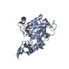
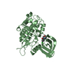
- PDB-4v1b: Structure of a novel carbohydrate binding module from glycoside h... -
+ Open data

- Basic information
| Entry | Database: PDB / ID: 4v1b | ||||||
|---|---|---|---|---|---|---|---|
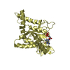
| Title | Structure of a novel carbohydrate binding module from glycoside hydrolase family 5 glucanase from Ruminococcus flavefaciens FD-1 collected at the Zn edge | ||||||
 Components Components | CARBOHYDRATE BINDING MODULE | ||||||
 Keywords Keywords | SUGAR BINDING PROTEIN / CELLULOSOME | ||||||
| Function / homology | Carbohydrate binding module Function and homology information Function and homology information | ||||||
| Biological species |  RUMINOCOCCUS FLAVEFACIENS (bacteria) RUMINOCOCCUS FLAVEFACIENS (bacteria) | ||||||
| Method |  X-RAY DIFFRACTION / SYNCHROTRON / MOLECULAR REPLACEMENT / Resolution: 2.69 Å X-RAY DIFFRACTION / SYNCHROTRON / MOLECULAR REPLACEMENT / Resolution: 2.69 Å | ||||||
 Authors Authors | Venditto, I. / Centeno, M.S.J. / Ferreira, L.M.A. / Fontes, C.M.G.A. / Najmudin, S. | ||||||
 Citation Citation | Journal: Proc.Natl.Acad.Sci.USA / Year: 2016 Title: Complexity of the Ruminococcus Flavefaciens Cellulosome Reflects an Expansion in Glycan Recognition. Authors: Venditto, I. / Luis, A.S. / Rydahl, M. / Schuckel, J. / Fernandes, V.O. / Vidal-Melgosa, S. / Bule, P. / Goyal, A. / Pires, V.M.R. / Dourado, C.G. / Ferreira, L.M.A. / Coutinho, P.M. / ...Authors: Venditto, I. / Luis, A.S. / Rydahl, M. / Schuckel, J. / Fernandes, V.O. / Vidal-Melgosa, S. / Bule, P. / Goyal, A. / Pires, V.M.R. / Dourado, C.G. / Ferreira, L.M.A. / Coutinho, P.M. / Henrissat, B. / Knox, J.P. / Basle, A. / Najmudin, S. / Gilbert, H.J. / Willats, W.G.T. / Fontes, C.M.G.A. #1: Journal: Acta Crystallogr.,Sect.F / Year: 2014 Title: Expression, Purification and Crystallization of a Novel Carbohydrate-Binding Module from the Ruminococcus Flavefaciens Cellulosome. Authors: Venditto, I. / Centeno, M.S.J. / Ferreira, L.M.A. / Fontes, C.M.G.A. / Najmudin, S. | ||||||
| History |
|
- Structure visualization
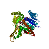
| Structure viewer | Molecule: MolmilJmol/JSmol |
|---|
- Downloads & links
-Download
| PDBx/mmCIF format | 4v1b.cif.gz | 126 KB | Display | PDBx/mmCIF format |
|---|---|---|---|---|
| PDB format | pdb4v1b.ent.gz | 101.3 KB | Display | PDB format |
| PDBx/mmJSON format | 4v1b.json.gz | Tree view | PDBx/mmJSON format | |
| Others |  Other downloads Other downloads |
-Validation report
| Arichive directory | https://data.pdbj.org/pub/pdb/validation_reports/v1/4v1bftp://data.pdbj.org/pub/pdb/validation_reports/v1/4v1b | HTTPS FTP |
|---|
-Related structure data
| Related structure data | 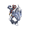 4d3lC 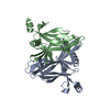 4v17C 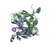 4v18SC 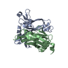 4v1iC 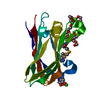 4v1kC 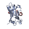 4v1lC 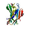 5aosC 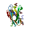 5aotC 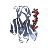 5fu2C  5fu3C  5fu4C 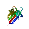 5fu5C C: citing same article ( S: Starting model for refinement |
|---|---|
| Similar structure data |


-Links
 PDBj
PDBj- Assembly
| Deposited unit | 
| ||||||||
|---|---|---|---|---|---|---|---|---|---|
| 1 |
| ||||||||
| Unit cell |
|
-Components
| #1: Protein | Mass: 17273.186 Da / Num. of mol.: 2 Source method: isolated from a genetically manipulated source Source: (gene. exp.) RUMINOCOCCUS FLAVEFACIENS (bacteria) / Strain: FD-1 / Production host: #2: Water | ChemComp-HOH / |  Mass: 18.015 Da / Num. of mol.: 4 / Source method: isolated from a natural source / Formula: H2O Mass: 18.015 Da / Num. of mol.: 4 / Source method: isolated from a natural source / Formula: H2OSequence details | GB WP009983134 | |
|---|
-Experimental details
-Experiment
| Experiment | Method: X-RAY DIFFRACTION / Number of used crystals: 1 |
|---|
- Sample preparation
| Crystal | Density Matthews: 2.57 Å3/Da / Density % sol: 52 % / Description: NONE |
|---|---|
| Crystal grow | pH: 7 Details: 0.2 M K2SO4, 20% PEG 3350. CRYO 30% GLYCEROL IN THE ABOVE CONDITION, PH 7.0 |
-Data collection
| Diffraction | Mean temperature: 100 K |
|---|---|
| Diffraction source | Source: SYNCHROTRON / Site: Diamond  / Beamline: I02 / Wavelength: 1.2855 / Beamline: I02 / Wavelength: 1.2855 |
| Detector | Type: DECTRIS PILATUS 6M / Detector: PIXEL / Date: May 6, 2013 |
| Radiation | Protocol: SINGLE WAVELENGTH / Monochromatic (M) / Laue (L): M / Scattering type: x-ray |
| Radiation wavelength | Wavelength: 1.2855 Å / Relative weight: 1 |
| Reflection | Resolution: 2.69→66.13 Å / Num. obs: 9947 / % possible obs: 99.8 % / Observed criterion σ(I): 0 / Redundancy: 19.3 % / Rmerge(I) obs: 0.15 / Net I/σ(I): 14.1 |
| Reflection shell | Resolution: 2.69→2.76 Å / Redundancy: 19 % / Rmerge(I) obs: 1.5 / Mean I/σ(I) obs: 3.2 / % possible all: 99.7 |
- Processing
| Software |
| ||||||||||||||||||||||||||||||||||||||||||||||||||||||||||||||||||||||||||||||||||||||||||||||||||||||||||||||||||||||||||||||||||||||||||||||||||||||||||||||||||||||||||||||||||||||
|---|---|---|---|---|---|---|---|---|---|---|---|---|---|---|---|---|---|---|---|---|---|---|---|---|---|---|---|---|---|---|---|---|---|---|---|---|---|---|---|---|---|---|---|---|---|---|---|---|---|---|---|---|---|---|---|---|---|---|---|---|---|---|---|---|---|---|---|---|---|---|---|---|---|---|---|---|---|---|---|---|---|---|---|---|---|---|---|---|---|---|---|---|---|---|---|---|---|---|---|---|---|---|---|---|---|---|---|---|---|---|---|---|---|---|---|---|---|---|---|---|---|---|---|---|---|---|---|---|---|---|---|---|---|---|---|---|---|---|---|---|---|---|---|---|---|---|---|---|---|---|---|---|---|---|---|---|---|---|---|---|---|---|---|---|---|---|---|---|---|---|---|---|---|---|---|---|---|---|---|---|---|---|---|
| Refinement | Method to determine structure: MOLECULAR REPLACEMENT Starting model: PDB ENTRY 4V18 Resolution: 2.69→66.13 Å / Cor.coef. Fo:Fc: 0.956 / Cor.coef. Fo:Fc free: 0.943 / SU B: 27.482 / SU ML: 0.241 / Cross valid method: THROUGHOUT / ESU R: 1.285 / ESU R Free: 0.273 / Stereochemistry target values: MAXIMUM LIKELIHOOD Details: HYDROGENS HAVE BEEN ADDED IN THE RIDING POSITIONS. U VALUES WITH TLS ADDED
| ||||||||||||||||||||||||||||||||||||||||||||||||||||||||||||||||||||||||||||||||||||||||||||||||||||||||||||||||||||||||||||||||||||||||||||||||||||||||||||||||||||||||||||||||||||||
| Solvent computation | Ion probe radii: 0.7 Å / Shrinkage radii: 0.7 Å / VDW probe radii: 1 Å / Solvent model: MASK | ||||||||||||||||||||||||||||||||||||||||||||||||||||||||||||||||||||||||||||||||||||||||||||||||||||||||||||||||||||||||||||||||||||||||||||||||||||||||||||||||||||||||||||||||||||||
| Displacement parameters | Biso mean: 80.606 Å2
| ||||||||||||||||||||||||||||||||||||||||||||||||||||||||||||||||||||||||||||||||||||||||||||||||||||||||||||||||||||||||||||||||||||||||||||||||||||||||||||||||||||||||||||||||||||||
| Refinement step | Cycle: LAST / Resolution: 2.69→66.13 Å
| ||||||||||||||||||||||||||||||||||||||||||||||||||||||||||||||||||||||||||||||||||||||||||||||||||||||||||||||||||||||||||||||||||||||||||||||||||||||||||||||||||||||||||||||||||||||
| Refine LS restraints |
|