 Movie
Movie Controller

[English] 日本語
 Yorodumi
- PDB-1wd0: Crystal structures of the hyperthermophilic chromosomal protein S... -
+ Open data

- Basic information
| Entry | Database: PDB / ID: 1wd0 | ||||||
|---|---|---|---|---|---|---|---|
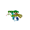
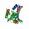
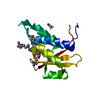
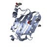
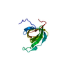
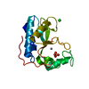
| Title | Crystal structures of the hyperthermophilic chromosomal protein Sac7d in complex with DNA decamers | ||||||
 Components Components |
| ||||||
 Keywords Keywords | structural protein/DNA / Protein-DNA complex / structural protein-DNA COMPLEX | ||||||
| Function / homology |  Function and homology information Function and homology information | ||||||
| Biological species |   Sulfolobus acidocaldarius (acidophilic) Sulfolobus acidocaldarius (acidophilic) | ||||||
| Method |  X-RAY DIFFRACTION / MOLECULAR REPLACEMENT / Resolution: 1.9 Å X-RAY DIFFRACTION / MOLECULAR REPLACEMENT / Resolution: 1.9 Å | ||||||
 Authors Authors | Ko, T.-P. / Chu, H.-M. / Chen, C.-Y. / Chou, C.-C. / Wang, A.H.-J. | ||||||
 Citation Citation | Journal: Acta Crystallogr.,Sect.D / Year: 2004 Title: Structures of the hyperthermophilic chromosomal protein Sac7d in complex with DNA decamers. Authors: Ko, T.P. / Chu, H.M. / Chen, C.Y. / Chou, C.C. / Wang, A.H. | ||||||
| History |
|
- Structure visualization
| Structure viewer | Molecule: MolmilJmol/JSmol |
|---|
- Downloads & links
-Download
| PDBx/mmCIF format | 1wd0.cif.gz | 40 KB | Display | PDBx/mmCIF format |
|---|---|---|---|---|
| PDB format | pdb1wd0.ent.gz | 25.4 KB | Display | PDB format |
| PDBx/mmJSON format | 1wd0.json.gz | Tree view | PDBx/mmJSON format | |
| Others |  Other downloads Other downloads |
-Validation report
| Arichive directory | https://data.pdbj.org/pub/pdb/validation_reports/wd/1wd0ftp://data.pdbj.org/pub/pdb/validation_reports/wd/1wd0 | HTTPS FTP |
|---|
-Related structure data
| Related structure data |  1wd1C  1azpS S: Starting model for refinement C: citing same article ( |
|---|---|
| Similar structure data |


-Links
 PDBj
PDBj- Assembly
| Deposited unit | 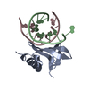
| ||||||||
|---|---|---|---|---|---|---|---|---|---|
| 1 |
| ||||||||
| Unit cell |
| ||||||||
| Details | monomer Sac7d is bound to DNA, DNA forms virtual double helices |
-Components
| #1: DNA chain | Mass: 3044.016 Da / Num. of mol.: 2 / Source method: obtained synthetically #2: Protein | | Mass: 7626.914 Da / Num. of mol.: 1 Source method: isolated from a genetically manipulated source Source: (gene. exp.) Sulfolobus acidocaldarius (acidophilic)Plasmid: pET-3b / Species (production host): Escherichia coli / Production host:  #3: Water | ChemComp-HOH / |  Mass: 18.015 Da / Num. of mol.: 95 / Source method: isolated from a natural source / Formula: H2O Mass: 18.015 Da / Num. of mol.: 95 / Source method: isolated from a natural source / Formula: H2O |
|---|
-Experimental details
-Experiment
| Experiment | Method: X-RAY DIFFRACTION / Number of used crystals: 1 |
|---|
- Sample preparation
| Crystal | Density Matthews: 1.8 Å3/Da / Density % sol: 30 % | ||||||||||||||||||||||||||||
|---|---|---|---|---|---|---|---|---|---|---|---|---|---|---|---|---|---|---|---|---|---|---|---|---|---|---|---|---|---|
| Crystal grow | Temperature: 298 K / Method: vapor diffusion, hanging drop / pH: 6.5 Details: Tris-HCl, PEG400, pH 6.5, VAPOR DIFFUSION, HANGING DROP, temperature 298K | ||||||||||||||||||||||||||||
| Components of the solutions |
|
-Data collection
| Diffraction | Mean temperature: 100 K |
|---|---|
| Diffraction source | Source: ROTATING ANODE / Type: RIGAKU MICROMAX-002 / Wavelength: 1.5418 Å |
| Detector | Type: RIGAKU RAXIS IV / Detector: IMAGE PLATE / Date: Feb 7, 2003 |
| Radiation | Monochromator: Ni FILTER / Protocol: SINGLE WAVELENGTH / Monochromatic (M) / Laue (L): M / Scattering type: x-ray |
| Radiation wavelength | Wavelength: 1.5418 Å / Relative weight: 1 |
| Reflection | Resolution: 1.9→30 Å / Num. all: 8617 / Num. obs: 8591 / % possible obs: 99.7 % / Observed criterion σ(F): 0 / Observed criterion σ(I): 0 / Redundancy: 6.5 % / Rmerge(I) obs: 0.045 / Net I/σ(I): 43 |
| Reflection shell | Resolution: 1.9→1.97 Å / Redundancy: 4.1 % / Rmerge(I) obs: 0.49 / Mean I/σ(I) obs: 2.3 / Num. unique all: 827 / % possible all: 97.5 |
- Processing
| Software |
| |||||||||||||||||||||||||
|---|---|---|---|---|---|---|---|---|---|---|---|---|---|---|---|---|---|---|---|---|---|---|---|---|---|---|
| Refinement | Method to determine structure: MOLECULAR REPLACEMENT Starting model: pdb 1azp Resolution: 1.9→30 Å / Isotropic thermal model: isotropic / Cross valid method: THROUGHOUT / σ(F): 0 / Stereochemistry target values: Engh & Huber
| |||||||||||||||||||||||||
| Displacement parameters | Biso mean: 38.5 Å2
| |||||||||||||||||||||||||
| Refine analyze |
| |||||||||||||||||||||||||
| Refinement step | Cycle: LAST / Resolution: 1.9→30 Å
| |||||||||||||||||||||||||
| Refine LS restraints |
| |||||||||||||||||||||||||
| LS refinement shell | Resolution: 1.9→1.97 Å / Rfactor Rfree error: 0.017
|
