- PDB-1w3s: The crystal structure of RecO from Deinococcus radiodurans. -
+
Open data
ID or keywords:
Loading...
-
Basic information
Entry
Database: PDB / ID: 1w3s
Title
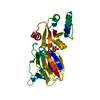
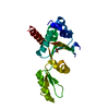
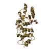
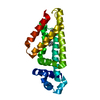
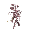
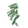
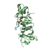
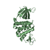
The crystal structure of RecO from Deinococcus radiodurans.
Components
HYPOTHETICAL PROTEIN DR0819
Keywords
DNA REPAIR / SAD / CYS4 ZINC FINGER / OB-FOLD / RECO
Function / homology
Function and homology information
bacterial nucleoid / double-strand break repair / DNA recombination / metal ion binding Similarity search - Function
Recombination protein O, zinc-binding domain / Recombination protein O, C-terminal domain / Erythroid Transcription Factor GATA-1; Chain A / Recombination protein O, RecO / DNA replication/recombination mediator RecO, N-terminal / Recombination protein O, C-terminal / Recombination protein O C terminal / Recombination protein O N terminal / ARFGAP/RecO-like zinc finger / de novo design (two linked rop proteins) ...Recombination protein O, zinc-binding domain / Recombination protein O, C-terminal domain / Erythroid Transcription Factor GATA-1; Chain A / Recombination protein O, RecO / DNA replication/recombination mediator RecO, N-terminal / Recombination protein O, C-terminal / Recombination protein O C terminal / Recombination protein O N terminal / ARFGAP/RecO-like zinc finger / de novo design (two linked rop proteins) / Other non-globular / Nucleic acid-binding proteins / Special / OB fold (Dihydrolipoamide Acetyltransferase, E2P) / Nucleic acid-binding, OB-fold / Up-down Bundle / Beta Barrel / Mainly Beta / Mainly Alpha Similarity search - Domain/homology
SHEET DETERMINATION METHOD: DSSP THE SHEETS PRESENTED AS "AA" AND "BA" IN EACH CHAIN ON SHEET ... SHEET DETERMINATION METHOD: DSSP THE SHEETS PRESENTED AS "AA" AND "BA" IN EACH CHAIN ON SHEET RECORDS BELOW ARE ACTUALLY 6-STRANDED BARRELS REPRESENTED BY A 7-STRANDED SHEET IN WHICH THE FIRST AND LAST STRANDS ARE IDENTICAL.
Mass: 18.015 Da / Num. of mol.: 23 / Source method: isolated from a natural source / Formula: H2O
-
Experimental details
-
Experiment
Experiment
Method: X-RAY DIFFRACTION / Number of used crystals: 1
-
Sample preparation
Crystal
Density Matthews: 2.9 Å3/Da / Density % sol: 58 % Description: THE STRUCTURE WAS SOLVED BY THE SAD METHOD ON A DATA SET COLLECTED AT THE ZINC ABSORPTION EDGE.
Protocol: SINGLE WAVELENGTH / Monochromatic (M) / Laue (L): M / Scattering type: x-ray
Radiation wavelength
Wavelength: 0.934 Å / Relative weight: 1
Reflection
Resolution: 2.4→48.6 Å / Num. obs: 26328 / % possible obs: 98.8 % / Observed criterion σ(I): 0 / Redundancy: 2.3 % / Rmerge(I) obs: 0.06 / Net I/σ(I): 9.6
Reflection shell
Resolution: 2.4→2.53 Å / Redundancy: 2.3 % / Rmerge(I) obs: 0.55 / Mean I/σ(I) obs: 1.4 / % possible all: 99.7
-
Processing
Software
Name
Version
Classification
XDS
datareduction
XDS
datascaling
SOLVE
phasing
SHARP
phasing
REFMAC
5.2.0003
refinement
Refinement
Method to determine structure: OTHER / Resolution: 2.4→95.35 Å / Cor.coef. Fo:Fc: 0.943 / Cor.coef. Fo:Fc free: 0.921 / SU B: 9.753 / SU ML: 0.219 / Cross valid method: THROUGHOUT / ESU R: 0.317 / ESU R Free: 0.247 / Stereochemistry target values: MAXIMUM LIKELIHOOD / Details: HYDROGENS HAVE BEEN ADDED IN THE RIDING POSITIONS.
Rfactor
Num. reflection
% reflection
Selection details
Rfree
0.267
1332
5.1 %
RANDOM
Rwork
0.225
-
-
-
obs
0.227
24995
98.5 %
-
Solvent computation
Ion probe radii: 0.8 Å / Shrinkage radii: 0.8 Å / VDW probe radii: 1.2 Å / Solvent model: BABINET MODEL WITH MASK
 Movie
Movie Controller


 Open data
 Basic information
 Components
 Keywords
 Function and homology information
 DEINOCOCCUS RADIODURANS (radioresistant)
 X-RAY DIFFRACTION /
  Authors
 Citation
 Structure visualization
 Downloads & links
 Other downloads


 PDBj
PDBj Assembly


 Mass: 65.409 Da / Num. of mol.: 2 / Source method: obtained synthetically / Formula: Zn
 Mass: 18.015 Da / Num. of mol.: 23 / Source method: isolated from a natural source / Formula: H2O
 Sample preparation
 / Beamline: ID14-1 / Wavelength: 0.934
  Processing