 Movie
Movie Controller

[English] 日本語
 Yorodumi
- PDB-3ig0: crystal structure of the second part of the Mycobacterium tubercu... -
+ Open data

- Basic information
| Entry | Database: PDB / ID: 3ig0 | ||||||
|---|---|---|---|---|---|---|---|
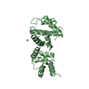
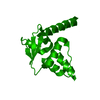
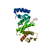
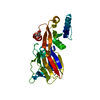
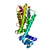
| Title | crystal structure of the second part of the Mycobacterium tuberculosis DNA gyrase reaction core: the TOPRIM domain at 2.1 A resolution | ||||||
 Components Components | DNA gyrase subunit B | ||||||
 Keywords Keywords | ISOMERASE / DNA gyrase / GyrB / TOPRIM / Type II topoisomerase / tuberculosis / quinolone binding site / DNA binding site / ATP-binding / Nucleotide-binding / Topoisomerase | ||||||
| Function / homology |  Function and homology information Function and homology informationDNA negative supercoiling activity / DNA topoisomerase type II (double strand cut, ATP-hydrolyzing) activity / DNA topoisomerase (ATP-hydrolysing) / DNA topological change / peptidoglycan-based cell wall / DNA-templated DNA replication / chromosome / response to antibiotic / magnesium ion binding / DNA binding ...DNA negative supercoiling activity / DNA topoisomerase type II (double strand cut, ATP-hydrolyzing) activity / DNA topoisomerase (ATP-hydrolysing) / DNA topological change / peptidoglycan-based cell wall / DNA-templated DNA replication / chromosome / response to antibiotic / magnesium ion binding / DNA binding / ATP binding / metal ion binding / plasma membrane / cytoplasm Similarity search - Function | ||||||
| Biological species |   Mycobacterium tuberculosis (bacteria) Mycobacterium tuberculosis (bacteria) | ||||||
| Method |  X-RAY DIFFRACTION / SYNCHROTRON / MOLECULAR REPLACEMENT / Resolution: 2.1 Å X-RAY DIFFRACTION / SYNCHROTRON / MOLECULAR REPLACEMENT / Resolution: 2.1 Å | ||||||
 Authors Authors | Piton, J. / Aubry, A. / Delarue, M. / Mayer, C. | ||||||
 Citation Citation | Journal: Plos One / Year: 2010 Title: Structural insights into the quinolone resistance mechanism of Mycobacterium tuberculosis DNA gyrase. Authors: Piton, J. / Petrella, S. / Delarue, M. / Andre-Leroux, G. / Jarlier, V. / Aubry, A. / Mayer, C. | ||||||
| History |
|
- Structure visualization
| Structure viewer | Molecule: MolmilJmol/JSmol |
|---|
- Downloads & links
-Download
| PDBx/mmCIF format | 3ig0.cif.gz | 53.6 KB | Display | PDBx/mmCIF format |
|---|---|---|---|---|
| PDB format | pdb3ig0.ent.gz | 36.9 KB | Display | PDB format |
| PDBx/mmJSON format | 3ig0.json.gz | Tree view | PDBx/mmJSON format | |
| Others |  Other downloads Other downloads |
-Validation report
| Arichive directory | https://data.pdbj.org/pub/pdb/validation_reports/ig/3ig0ftp://data.pdbj.org/pub/pdb/validation_reports/ig/3ig0 | HTTPS FTP |
|---|
-Related structure data
| Related structure data |  3ifzC 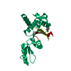 3m4iC 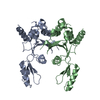 2zjtS S: Starting model for refinement C: citing same article ( |
|---|---|
| Similar structure data |


-Links
 PDBj
PDBj


- Assembly
| Deposited unit | 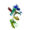
| ||||||||
|---|---|---|---|---|---|---|---|---|---|
| 1 |
| ||||||||
| Unit cell |
|
-Components
| #1: Protein | Mass: 27419.260 Da / Num. of mol.: 1 / Fragment: TOPRIM domain (C-terminal domain) Source method: isolated from a genetically manipulated source Source: (gene. exp.) Mycobacterium tuberculosis (bacteria) / Strain: HR37RV / Gene: gyrB, MT0005, MTCY10H4.03, Rv0005 / Plasmid: pRSF-2 Ek/LIC / Production host: References: UniProt: P0C5C5, UniProt: P9WG45*PLUS, EC: 5.99.1.3 |
|---|---|
| #2: Water | ChemComp-HOH /  Mass: 18.015 Da / Num. of mol.: 107 / Source method: isolated from a natural source / Formula: H2O Mass: 18.015 Da / Num. of mol.: 107 / Source method: isolated from a natural source / Formula: H2O |
-Experimental details
-Experiment
| Experiment | Method: X-RAY DIFFRACTION / Number of used crystals: 1 |
|---|
- Sample preparation
| Crystal | Density Matthews: 2.42 Å3/Da / Density % sol: 49.26 % |
|---|---|
| Crystal grow | Temperature: 293 K / Method: vapor diffusion, hanging drop / pH: 8 Details: ammonium sulfate, Tris, PEG 4000, Mgcl2, pH 8.0, VAPOR DIFFUSION, HANGING DROP, temperature 293K |
-Data collection
| Diffraction | Mean temperature: 100 K |
|---|---|
| Diffraction source | Source: SYNCHROTRON / Site: ESRF  / Beamline: ID23-1 / Wavelength: 0.9762 Å / Beamline: ID23-1 / Wavelength: 0.9762 Å |
| Detector | Type: ADSC QUANTUM 315r / Detector: CCD / Date: Jul 9, 2009 / Details: toroidal mirror + Si(111) monochromator |
| Radiation | Monochromator: Si(111) channel-cut / Protocol: SINGLE WAVELENGTH / Monochromatic (M) / Laue (L): M / Scattering type: x-ray |
| Radiation wavelength | Wavelength: 0.9762 Å / Relative weight: 1 |
| Reflection | Resolution: 2.1→13.96 Å / Num. all: 16640 / Num. obs: 16487 / % possible obs: 99.1 % / Observed criterion σ(F): 0 / Observed criterion σ(I): -3 / Redundancy: 8.6 % / Biso Wilson estimate: 33.746 Å2 / Rmerge(I) obs: 0.147 / Rsym value: 0.141 / Net I/σ(I): 13.1 |
| Reflection shell | Resolution: 2.1→2.3 Å / Redundancy: 4 % / Rmerge(I) obs: 0.556 / Mean I/σ(I) obs: 3.3 / Num. unique all: 3347 / Rsym value: 0.556 / % possible all: 86.5 |
- Processing
| Software |
| ||||||||||||||||||||||||||||||||||||||||||||||||||
|---|---|---|---|---|---|---|---|---|---|---|---|---|---|---|---|---|---|---|---|---|---|---|---|---|---|---|---|---|---|---|---|---|---|---|---|---|---|---|---|---|---|---|---|---|---|---|---|---|---|---|---|
| Refinement | Method to determine structure: MOLECULAR REPLACEMENT Starting model: 2ZJT Resolution: 2.1→13.96 Å / Isotropic thermal model: isotropic / Cross valid method: THROUGHOUT / σ(F): 0 / Stereochemistry target values: Engh & Huber
| ||||||||||||||||||||||||||||||||||||||||||||||||||
| Displacement parameters | Biso mean: 40.02 Å2
| ||||||||||||||||||||||||||||||||||||||||||||||||||
| Refine analyze | Luzzati coordinate error obs: 0.2861 Å | ||||||||||||||||||||||||||||||||||||||||||||||||||
| Refinement step | Cycle: LAST / Resolution: 2.1→13.96 Å
| ||||||||||||||||||||||||||||||||||||||||||||||||||
| Refine LS restraints |
| ||||||||||||||||||||||||||||||||||||||||||||||||||
| LS refinement shell | Resolution: 2.1→2.25 Å / Total num. of bins used: 8
|
