 Movie
Movie Controller

[English] 日本語
 Yorodumi
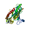
- PDB-1vzz: CRYSTAL STRUCTURE OF MUTANT ENZYME Y32F/D103L OF KETOSTEROID ISOM... -
+ Open data

- Basic information
| Entry | Database: PDB / ID: 1vzz | ||||||
|---|---|---|---|---|---|---|---|
| Title | CRYSTAL STRUCTURE OF MUTANT ENZYME Y32F/D103L OF KETOSTEROID ISOMERASE FROM PSEUDOMONAS PUTIDA BIOTYPE B | ||||||
 Components Components | STEROID DELTA-ISOMERASE | ||||||
 Keywords Keywords | ISOMERASE / CONESHELL / CLOSED BARREL / CURVED B-SHEET | ||||||
| Function / homology |  Function and homology information Function and homology informationsteroid Delta-isomerase / steroid Delta-isomerase activity / steroid metabolic process Similarity search - Function | ||||||
| Biological species |  PSEUDOMONAS PUTIDA (bacteria) PSEUDOMONAS PUTIDA (bacteria) | ||||||
| Method |  X-RAY DIFFRACTION / SYNCHROTRON / MOLECULAR REPLACEMENT / Resolution: 2.3 Å X-RAY DIFFRACTION / SYNCHROTRON / MOLECULAR REPLACEMENT / Resolution: 2.3 Å | ||||||
 Authors Authors | Jang, D.S. / Cha, H.J. / Choi, K.Y. | ||||||
 Citation Citation | Journal: Biochem.J. / Year: 2004 Title: Structural Double-Mutant Cycle Analysis of a Hydrogen Bond Network in Ketosteroid Isomerase from Pseudomonas Putida Biotype B. Authors: Jang, D.S. / Cha, H.J. / Cha, S.S. / Hong, B.H. / Ha, N.C. / Lee, J.Y. / Oh, B.H. / Lee, H.S. / Choi, K.Y. #1: Journal: Biochemistry / Year: 2000Title: Contribution of the Hydrogen Bond Network Involving a Tyrosine Triad in the Active Site to Structure and Function of a Highly Proficient Ketosteroid Isomerase from Pseudomonas Putida Biotype B Authors: Kim, D.H. / Jang, D.S. / Nam, G.H. / Choi, G.D. / Kim, J.S. / Ha, N.C. / Kim, M.S. / Oh, B.H. / Choi, K.Y. | ||||||
| History |
| ||||||
| Remark 700 | SHEET THE SHEET STRUCTURE OF THIS MOLECULE IS BIFURCATED. IN ORDER TO REPRESENT THIS FEATURE IN ... SHEET THE SHEET STRUCTURE OF THIS MOLECULE IS BIFURCATED. IN ORDER TO REPRESENT THIS FEATURE IN THE SHEET RECORDS BELOW, TWO SHEETS ARE DEFINED. |
- Structure visualization
| Structure viewer | Molecule: MolmilJmol/JSmol |
|---|
- Downloads & links
-Download
| PDBx/mmCIF format | 1vzz.cif.gz | 60.6 KB | Display | PDBx/mmCIF format |
|---|---|---|---|---|
| PDB format | pdb1vzz.ent.gz | 44.5 KB | Display | PDB format |
| PDBx/mmJSON format | 1vzz.json.gz | Tree view | PDBx/mmJSON format | |
| Others |  Other downloads Other downloads |
-Validation report
| Summary document | 1vzz_validation.pdf.gz | 416 KB | Display | wwPDB validaton report |
|---|---|---|---|---|
| Full document | 1vzz_full_validation.pdf.gz | 422.7 KB | Display | |
| Data in XML | 1vzz_validation.xml.gz | 10.8 KB | Display | |
| Data in CIF | 1vzz_validation.cif.gz | 14.7 KB | Display | |
| Arichive directory | https://data.pdbj.org/pub/pdb/validation_reports/vz/1vzzftp://data.pdbj.org/pub/pdb/validation_reports/vz/1vzz | HTTPS FTP |
-Related structure data
| Related structure data |  1w00C  1w01C  1w02C  1dmqS S: Starting model for refinement C: citing same article ( |
|---|---|
| Similar structure data |


-Links
 PDBj
PDBj
- Assembly
| Deposited unit | 
| ||||||||
|---|---|---|---|---|---|---|---|---|---|
| 1 |
| ||||||||
| Unit cell |
|
-Components
| #1: Protein | Mass: 14530.571 Da / Num. of mol.: 2 / Mutation: YES Source method: isolated from a genetically manipulated source Source: (gene. exp.) PSEUDOMONAS PUTIDA (bacteria) / Plasmid: PKK223-3 / Production host: #2: Water | ChemComp-HOH / |  Mass: 18.015 Da / Num. of mol.: 88 / Source method: isolated from a natural source / Formula: H2O Mass: 18.015 Da / Num. of mol.: 88 / Source method: isolated from a natural source / Formula: H2OCompound details | ENGINEERED | |
|---|
-Experimental details
-Experiment
| Experiment | Method: X-RAY DIFFRACTION / Number of used crystals: 1 |
|---|
- Sample preparation
| Crystal | Density Matthews: 2.12 Å3/Da / Density % sol: 42.05 % |
|---|---|
| Crystal grow | pH: 4.6 / Details: SODIUM ACETATE, AMMONIUM ACETATE, pH 4.60 |
-Data collection
| Diffraction | Mean temperature: 100 K |
|---|---|
| Diffraction source | Source: SYNCHROTRON / Site: Photon Factory  / Beamline: BL-6B / Wavelength: 1.5418 / Beamline: BL-6B / Wavelength: 1.5418 |
| Detector | Type: MACSCIENCE / Detector: IMAGE PLATE / Date: Mar 5, 2003 |
| Radiation | Protocol: SINGLE WAVELENGTH / Monochromatic (M) / Laue (L): M / Scattering type: x-ray |
| Radiation wavelength | Wavelength: 1.5418 Å / Relative weight: 1 |
| Reflection | Resolution: 2.15→20 Å / Num. obs: 6746 / % possible obs: 91.7 % / Observed criterion σ(I): 1 / Redundancy: 6.9 % / Biso Wilson estimate: 13.8 Å2 / Rmerge(I) obs: 0.053 / Net I/σ(I): 6.3 |
| Reflection shell | Resolution: 2.15→2.25 Å / Redundancy: 3.8 % / Rmerge(I) obs: 0.226 / % possible all: 82.3 |
- Processing
| Software |
| ||||||||||||||||||||||||||||||||||||||||||||||||||||||||||||
|---|---|---|---|---|---|---|---|---|---|---|---|---|---|---|---|---|---|---|---|---|---|---|---|---|---|---|---|---|---|---|---|---|---|---|---|---|---|---|---|---|---|---|---|---|---|---|---|---|---|---|---|---|---|---|---|---|---|---|---|---|---|
| Refinement | Method to determine structure: MOLECULAR REPLACEMENT Starting model: PDB ENTRY 1DMQ Resolution: 2.3→28.41 Å / Rfactor Rfree error: 0.007 / Data cutoff high absF: 2640441.64 / Isotropic thermal model: RESTRAINED / Cross valid method: THROUGHOUT / σ(F): 0
| ||||||||||||||||||||||||||||||||||||||||||||||||||||||||||||
| Solvent computation | Solvent model: FLAT MODEL / ksol: 0.379582 e/Å3 | ||||||||||||||||||||||||||||||||||||||||||||||||||||||||||||
| Displacement parameters | Biso mean: 20.7 Å2
| ||||||||||||||||||||||||||||||||||||||||||||||||||||||||||||
| Refine analyze |
| ||||||||||||||||||||||||||||||||||||||||||||||||||||||||||||
| Refinement step | Cycle: LAST / Resolution: 2.3→28.41 Å
| ||||||||||||||||||||||||||||||||||||||||||||||||||||||||||||
| Refine LS restraints |
| ||||||||||||||||||||||||||||||||||||||||||||||||||||||||||||
| LS refinement shell | Resolution: 2.3→2.44 Å / Rfactor Rfree error: 0.019 / Total num. of bins used: 6
| ||||||||||||||||||||||||||||||||||||||||||||||||||||||||||||
| Xplor file |
|
