 Movie
Movie Controller

[English] 日本語
 Yorodumi
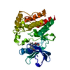
- PDB-1e3r: Crystal structure of ketosteroid isomerase mutant D40N (D38N TI n... -
+ Open data

- Basic information
| Entry | Database: PDB / ID: 1e3r | ||||||
|---|---|---|---|---|---|---|---|
| Title | Crystal structure of ketosteroid isomerase mutant D40N (D38N TI numbering) from Pseudomonas putida complexed with androsten-3beta-ol-17-one | ||||||
 Components Components | ISOMERASE | ||||||
 Keywords Keywords | ISOMERASE / KSI KETOSTEROIED ISOMERASE / LBHB / ANDROSTEN-3BETA-OL-17-ONE | ||||||
| Function / homology |  Function and homology information Function and homology informationsteroid Delta-isomerase / steroid Delta-isomerase activity / steroid metabolic process Similarity search - Function | ||||||
| Biological species |  PSEUDOMONAS PUTIDA (bacteria) PSEUDOMONAS PUTIDA (bacteria) | ||||||
| Method |  X-RAY DIFFRACTION / SYNCHROTRON / MOLECULAR REPLACEMENT / Resolution: 2.5 Å X-RAY DIFFRACTION / SYNCHROTRON / MOLECULAR REPLACEMENT / Resolution: 2.5 Å | ||||||
 Authors Authors | Ha, N.-C. / Kim, M.-S. / Hyun, B.-H. / Oh, B.-H. | ||||||
 Citation Citation | Journal: J.Biol.Chem. / Year: 2000 Title: Detection of Large Pka Perturbations of an Inhibitor and a Catalytic Group at an Enzyme Active Site, a Mechanistic Basis for Catalytic Power of Many Enzymes Authors: Ha, N.-C. / Kim, M.-S. / Lee, W. / Choi, K.Y. / Oh, B.-H. | ||||||
| History |
|
- Structure visualization
| Structure viewer | Molecule: MolmilJmol/JSmol |
|---|
- Downloads & links
-Download
| PDBx/mmCIF format | 1e3r.cif.gz | 60.5 KB | Display | PDBx/mmCIF format |
|---|---|---|---|---|
| PDB format | pdb1e3r.ent.gz | 44.8 KB | Display | PDB format |
| PDBx/mmJSON format | 1e3r.json.gz | Tree view | PDBx/mmJSON format | |
| Others |  Other downloads Other downloads |
-Validation report
| Arichive directory | https://data.pdbj.org/pub/pdb/validation_reports/e3/1e3rftp://data.pdbj.org/pub/pdb/validation_reports/e3/1e3r | HTTPS FTP |
|---|
-Related structure data


-Links
 PDBj
PDBj
- Assembly
| Deposited unit | 
| ||||||||
|---|---|---|---|---|---|---|---|---|---|
| 1 |
| ||||||||
| Unit cell |
|
-Components
| #1: Protein | Mass: 14547.515 Da / Num. of mol.: 2 / Mutation: YES Source method: isolated from a genetically manipulated source Details: ANDROSTENE-3BETA-OL-17-ONE / Source: (gene. exp.) PSEUDOMONAS PUTIDA (bacteria) / Production host: #2: Chemical |   Mass: 288.424 Da / Num. of mol.: 2 / Source method: obtained synthetically / Formula: C19H28O2 / Comment: hormone*YM Mass: 288.424 Da / Num. of mol.: 2 / Source method: obtained synthetically / Formula: C19H28O2 / Comment: hormone*YM#3: Water | ChemComp-HOH / |  Mass: 18.015 Da / Num. of mol.: 76 / Source method: isolated from a natural source / Formula: H2O Mass: 18.015 Da / Num. of mol.: 76 / Source method: isolated from a natural source / Formula: H2OCompound details | CHAIN A, B ENGINEERED | |
|---|
-Experimental details
-Experiment
| Experiment | Method: X-RAY DIFFRACTION / Number of used crystals: 1 |
|---|
- Sample preparation
| Crystal | Density Matthews: 2.7 Å3/Da / Density % sol: 54.43 % | ||||||||||||||||||||
|---|---|---|---|---|---|---|---|---|---|---|---|---|---|---|---|---|---|---|---|---|---|
| Crystal grow | pH: 6.5 / Details: pH 6.50 | ||||||||||||||||||||
| Crystal grow | *PLUS Temperature: 22 ℃ / pH: 4.6 / Method: vapor diffusion, hanging drop / Details: Kim, S.W., (1997) Biochemistry, 36, 14030. | ||||||||||||||||||||
| Components of the solutions | *PLUS
|
-Data collection
| Diffraction | Mean temperature: 292 K |
|---|---|
| Diffraction source | Source: SYNCHROTRON / Site: Photon Factory  / Beamline: BL-18B / Wavelength: 1 / Beamline: BL-18B / Wavelength: 1 |
| Detector | Type: FUJI / Detector: IMAGE PLATE / Details: MIRRORS |
| Radiation | Protocol: SINGLE WAVELENGTH / Monochromatic (M) / Laue (L): M / Scattering type: x-ray |
| Radiation wavelength | Wavelength: 1 Å / Relative weight: 1 |
| Reflection | Resolution: 2.5→20 Å / Num. obs: 11746 / % possible obs: 82.2 % / Observed criterion σ(I): 1 / Redundancy: 3 % / Rmerge(I) obs: 0.065 / Rsym value: 0.065 |
| Reflection | *PLUS Lowest resolution: 20 Å |
| Reflection shell | *PLUS % possible obs: 66.4 % |
- Processing
| Software |
| ||||||||||||||||||||||||||||||||||||||||||||||||||||||||||||
|---|---|---|---|---|---|---|---|---|---|---|---|---|---|---|---|---|---|---|---|---|---|---|---|---|---|---|---|---|---|---|---|---|---|---|---|---|---|---|---|---|---|---|---|---|---|---|---|---|---|---|---|---|---|---|---|---|---|---|---|---|---|
| Refinement | Method to determine structure: MOLECULAR REPLACEMENT Starting model: 1E3R Resolution: 2.5→8 Å / Cross valid method: THROUGHOUT / σ(F): 1
| ||||||||||||||||||||||||||||||||||||||||||||||||||||||||||||
| Refinement step | Cycle: LAST / Resolution: 2.5→8 Å
| ||||||||||||||||||||||||||||||||||||||||||||||||||||||||||||
| Refine LS restraints |
|
