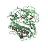
[English] 日本語
Yorodumi
- PDB-1vkz: Crystal structure of Phosphoribosylamine--glycine ligase (TM1250)... -

+
Open data


ID or keywords:

Loading...

-
Basic information

Entry
Database: PDB / ID: 1vkz
TitleCrystal structure of Phosphoribosylamine--glycine ligase (TM1250) from Thermotoga maritima at 2.30 A resolution
ComponentsPhosphoribosylamine--glycine ligase
KeywordsLIGASE / TM1250 / PHOSPHORIBOSYLAMINE--GLYCINE LIGASE / STRUCTURAL GENOMICS / JCSG / PROTEIN STRUCTURE INITIATIVE / PSI / Joint Center for Structural Genomics
Function / homology
Function and homology information


phosphoribosylamine-glycine ligase / phosphoribosylamine-glycine ligase activity / purine nucleobase biosynthetic process / 'de novo' IMP biosynthetic process / ATP binding / metal ion binding
Similarity search - Function
Phosphoribosylglycinamide synthetase, C-terminal domain / Glycinamide Ribonucleotide Synthetase; Chain A, domain 4 / Phosphoribosylglycinamide synthetase / Phosphoribosylglycinamide synthetase, conserved site / Phosphoribosylglycinamide synthetase, C-domain / Phosphoribosylglycinamide synthetase, N-terminal / Phosphoribosylglycinamide synthetase, C-domain superfamily / Phosphoribosylglycinamide synthetase, C domain / Phosphoribosylglycinamide synthetase, N domain / Phosphoribosylglycinamide synthetase signature. ...Phosphoribosylglycinamide synthetase, C-terminal domain / Glycinamide Ribonucleotide Synthetase; Chain A, domain 4 / Phosphoribosylglycinamide synthetase / Phosphoribosylglycinamide synthetase, conserved site / Phosphoribosylglycinamide synthetase, C-domain / Phosphoribosylglycinamide synthetase, N-terminal / Phosphoribosylglycinamide synthetase, C-domain superfamily / Phosphoribosylglycinamide synthetase, C domain / Phosphoribosylglycinamide synthetase, N domain / Phosphoribosylglycinamide synthetase signature. / Phosphoribosylglycinamide synthetase, C domain / Phosphoribosylglycinamide synthetase, ATP-grasp (A) domain / Phosphoribosylglycinamide synthetase, ATP-grasp (A) domain / Rossmann fold - #20 / Rudiment single hybrid motif / ATP-grasp fold, A domain / ATP-grasp fold, B domain / ATP-grasp fold, subdomain 1 / Pre-ATP-grasp domain superfamily / ATP-grasp fold / ATP-grasp fold profile. / D-amino Acid Aminotransferase; Chain A, domain 1 / Dna Ligase; domain 1 / Alpha-Beta Complex / Rossmann fold / 2-Layer Sandwich / 3-Layer(aba) Sandwich / Alpha Beta
Similarity search - Domain/homology
Phosphoribosylamine--glycine ligase
Similarity search - Component
Biological speciesThermotoga maritima (bacteria)
MethodX-RAY DIFFRACTION / SYNCHROTRON / MAD / Resolution: 2.3 Å
AuthorsJoint Center for Structural Genomics (JCSG)
CitationJournal: To be published
Title: Crystal structure of Phosphoribosylamine--glycine ligase (TM1250) from Thermotoga maritima at 2.30 A resolution
Authors: Joint Center for Structural Genomics (JCSG)
History
DepositionJun 29, 2004Deposition site: RCSB / Processing site: RCSB
Revision 1.0Aug 24, 2004Provider: repository / Type: Initial release
Revision 1.1Apr 26, 2008Group: Version format compliance
Revision 1.2Jul 13, 2011Group: Advisory / Version format compliance
Revision 1.3Jan 25, 2023Group: Database references / Derived calculations
Category: database_2 / struct_conn ...database_2 / struct_conn / struct_ref_seq_dif / struct_site
Item: _database_2.pdbx_DOI / _database_2.pdbx_database_accession ..._database_2.pdbx_DOI / _database_2.pdbx_database_accession / _struct_conn.pdbx_leaving_atom_flag / _struct_ref_seq_dif.details / _struct_site.pdbx_auth_asym_id / _struct_site.pdbx_auth_comp_id / _struct_site.pdbx_auth_seq_id

-
Structure visualization

Structure viewerMolecule:
MolmilJmol/JSmol

Downloads & links

-
Assembly

Deposited unit
A: Phosphoribosylamine--glycine ligase
B: Phosphoribosylamine--glycine ligase
hetero molecules


Theoretical massNumber of molelcules
Total (without water)92,6113
Polymers92,5492
Non-polymers621
Water2,180121
1
A: Phosphoribosylamine--glycine ligase


Theoretical massNumber of molelcules
Total (without water)46,2741
Polymers46,2741
Non-polymers00
Water181
TypeNameSymmetry operationNumber
identity operation1_555x,y,z1
2
B: Phosphoribosylamine--glycine ligase
hetero molecules


Theoretical massNumber of molelcules
Total (without water)46,3362
Polymers46,2741
Non-polymers621
Water181
TypeNameSymmetry operationNumber
identity operation1_555x,y,z1
Unit cell
Length a, b, c (Å)63.745, 78.443, 85.018
Angle α, β, γ (deg.)90.00, 95.06, 90.00
Int Tables number4
Space group name H-MP1211
Noncrystallographic symmetry (NCS)NCS domain:
IDEns-IDDetails
11A
21B
12A
22B
32A
42B
52A
62B
13A
23B
33A
43B
14A
24B
34A
44B

NCS domain segments:

Refine code: 2

Dom-IDComponent-IDEns-IDBeg label comp-IDEnd label comp-IDAuth asym-IDLabel asym-IDAuth seq-IDLabel seq-ID
111ALALEUAA110 - 180122 - 192
211ALALEUBB110 - 180122 - 192
112GLYTHRAA83 - 10995 - 121
212GLYTHRBB83 - 10995 - 121
322ALALEUAA181 - 199193 - 211
422ALALEUBB181 - 199193 - 211
532GLULEUAA227 - 278239 - 290
632GLULEUBB227 - 278239 - 290
113PROVALAA200 - 226212 - 238
213PROVALBB200 - 226212 - 238
323PHEALAAA315 - 399327 - 411
423PHEALABB315 - 399327 - 411
114VALPHEAA4 - 8216 - 94
214VALPHEBB4 - 8216 - 94
324GLYGLYAA279 - 314291 - 326
424GLYGLYBB279 - 314291 - 326

NCS ensembles :
ID
1
2
3
4

-
Components

#1: Protein Phosphoribosylamine--glycine ligase / GARS / Glycinamide ribonucleotide synthetase / Phosphoribosylglycinamide synthetase


Mass: 46274.418 Da / Num. of mol.: 2 / Mutation: 6.3.4.13
Source method: isolated from a genetically manipulated source
Source: (gene. exp.) Thermotoga maritima (bacteria) / Gene: purD, TM1250 / Production host: Escherichia coli (E. coli)
References: UniProt: Q9X0X7, phosphoribosylamine-glycine ligase
#2: Chemical ChemComp-EDO / 1,2-ETHANEDIOL / ETHYLENE GLYCOL / Ethylene glycol


Mass: 62.068 Da / Num. of mol.: 1 / Source method: obtained synthetically / Formula: C2H6O2
#3: Water ChemComp-HOH / water / Water


Mass: 18.015 Da / Num. of mol.: 121 / Source method: isolated from a natural source / Formula: H2O

-
Experimental details

-
Experiment

ExperimentMethod: X-RAY DIFFRACTION / Number of used crystals: 2

-
Sample preparation

Crystal
IDDensity Matthews3/Da)Density % sol (%)Description
12.2946.21
22.4449.26TWO CRYSTALS WERE USED IN THE SOLUTION OF THE STRUCTURE. ONE CRYSTAL WAS USED FOR SE-MET MAD PHASING AND ANOTHER WAS USED FOR PHASE EXTENSION AND REFINEMENT.
Crystal grow
Temperature (K)Crystal-IDMethodpHDetails
2771vapor diffusion, sitting drop, nanodrop6.518% PEG-8000, 0.1M sodium cacodylate pH 6.5, 0.2M calcium acetate hydrate, VAPOR DIFFUSION,SITTING DROP,NANODROP, temperature 277K
2772vapor diffusion, sitting drop, nanodrop720% PEG-6000, 0.1M HEPES pH 7.0, VAPOR DIFFUSION,SITTING DROP,NANODROP, temperature 277K

-
Data collection

Diffraction
IDMean temperature (K)Crystal-ID
11001
21001
1,21
Diffraction source
SourceSiteBeamlineIDWavelengthWavelength (Å)
SYNCHROTRONALS 8.2.111
SYNCHROTRONAPS 19-BM20.979127,0.979292,0.964063
Detector
TypeIDDetectorDateDetails
ADSC1CCDMar 3, 2004
APS 2CCDFeb 20, 2004water cooled, sagitally focusing 2nd crystal, Rosenbaum-Rock vertical focusing mirror
Radiation
IDMonochromatorProtocolMonochromatic (M) / Laue (L)Scattering typeWavelength-ID
1Double Crystal Si(111)SINGLE WAVELENGTHMx-ray1
2Rosenbaum-Rock double-crystal monochromatorMADMx-ray2
Radiation wavelength
IDWavelength (Å)Relative weight
111
20.9791271
30.9792921
40.9640631
ReflectionResolution: 2.3→37.4 Å / Num. obs: 31749 / % possible obs: 85.6 % / Redundancy: 2 % / Biso Wilson estimate: 54.3 Å2 / Rmerge(I) obs: 0.054 / Net I/σ(I): 12.1
Reflection shellResolution: 2.3→2.36 Å / Redundancy: 1.9 % / Rmerge(I) obs: 0.632 / Mean I/σ(I) obs: 1.3 / Num. unique all: 2434 / % possible all: 88.2

-
Processing

Software
NameVersionClassification
DENZOdata reduction
SCALA4.2)data scaling
SOLVEphasing
REFMAC5.2.0000refinement
CCP4(SCALA)data scaling
RefinementMethod to determine structure: MAD / Resolution: 2.3→37.26 Å / Cor.coef. Fo:Fc: 0.957 / Cor.coef. Fo:Fc free: 0.929 / SU B: 20.438 / SU ML: 0.232 / TLS residual ADP flag: LIKELY RESIDUAL / Cross valid method: THROUGHOUT / ESU R: 0.591 / ESU R Free: 0.299
Stereochemistry target values: MAXIMUM LIKELIHOOD WITH PHASES
Details: HYDROGENS HAVE BEEN ADDED IN THE RIDING POSITIONS. THE PHASES USED FOR THE RESTRAINTS WERE DENSITY MODIFIED (SOLVENT FLATTENED, HISTOGRAM, MULTI RESOLUTION) PHASES EXTENDED FROM STARTING MAD ...Details: HYDROGENS HAVE BEEN ADDED IN THE RIDING POSITIONS. THE PHASES USED FOR THE RESTRAINTS WERE DENSITY MODIFIED (SOLVENT FLATTENED, HISTOGRAM, MULTI RESOLUTION) PHASES EXTENDED FROM STARTING MAD PHASES FROM A DIFFERENT CRYSTAL. THE FOLLOWING RESIDUES HAVE UNMODELED ATOMS DUE TO MISSING DENSITY: A141 A151 A156 A303 A340 A374 A378 B54 B86 B141 B151 B303. NO DENSITY WAS PRESENT FOR THE LOOP EXTENDING FROM RESIDUES 73 TO 77.
RfactorNum. reflection% reflectionSelection details
Rfree0.26383 1560 5 %RANDOM
Rwork0.21315 ---
obs0.21572 29714 84.01 %-
Solvent computationIon probe radii: 0.8 Å / Shrinkage radii: 0.8 Å / VDW probe radii: 1.2 Å / Solvent model: BABINET MODEL WITH MASK
Displacement parametersBiso mean: 44.622 Å2
Baniso -1Baniso -2Baniso -3
1-0.3 Å20 Å2-0.69 Å2
2--2.77 Å20 Å2
3----3.19 Å2
Refinement stepCycle: LAST / Resolution: 2.3→37.26 Å
ProteinNucleic acidLigandSolventTotal
Num. atoms6065 0 4 121 6190
Refine LS restraints
Refine-IDTypeDev idealDev ideal targetNumber
X-RAY DIFFRACTIONr_bond_refined_d0.0160.0226198
X-RAY DIFFRACTIONr_bond_other_d0.0020.025742
X-RAY DIFFRACTIONr_angle_refined_deg1.4911.9668355
X-RAY DIFFRACTIONr_angle_other_deg0.811313357
X-RAY DIFFRACTIONr_dihedral_angle_1_deg7.2935778
X-RAY DIFFRACTIONr_dihedral_angle_2_deg32.51723.723274
X-RAY DIFFRACTIONr_dihedral_angle_3_deg17.106151078
X-RAY DIFFRACTIONr_dihedral_angle_4_deg19.7571543
X-RAY DIFFRACTIONr_chiral_restr0.0830.2890
X-RAY DIFFRACTIONr_gen_planes_refined0.0050.026921
X-RAY DIFFRACTIONr_gen_planes_other0.0010.021264
X-RAY DIFFRACTIONr_nbd_refined0.2070.21337
X-RAY DIFFRACTIONr_nbd_other0.1830.26078
X-RAY DIFFRACTIONr_nbtor_other0.0890.24016
X-RAY DIFFRACTIONr_xyhbond_nbd_refined0.1770.2224
X-RAY DIFFRACTIONr_symmetry_vdw_refined0.1810.220
X-RAY DIFFRACTIONr_symmetry_vdw_other0.220.259
X-RAY DIFFRACTIONr_symmetry_hbond_refined0.1430.28
X-RAY DIFFRACTIONr_mcbond_it1.93734146
X-RAY DIFFRACTIONr_mcbond_other0.47931624
X-RAY DIFFRACTIONr_mcangle_it2.7656161
X-RAY DIFFRACTIONr_scbond_it5.18482578
X-RAY DIFFRACTIONr_scangle_it7.017112194
X-RAY DIFFRACTIONr_xyhbond_nbd_other0.0570.21
Refine LS restraints NCS

Dom-ID: 1 / Auth asym-ID: A / Refine-ID: X-RAY DIFFRACTION

Ens-IDNumberTypeRms dev position (Å)Weight position
1415tight positional0.030.05
1670medium positional0.320.5
1415tight thermal0.090.5
1670medium thermal0.512
2575tight positional0.030.05
2967medium positional0.240.5
2575tight thermal0.140.5
2967medium thermal0.72
3650tight positional0.030.05
3998medium positional0.360.5
3650tight thermal0.120.5
3998medium thermal0.652
4635tight positional0.040.05
4966medium positional0.360.5
4635tight thermal0.120.5
4966medium thermal0.692
LS refinement shellResolution: 2.3→2.36 Å / Total num. of bins used: 20
RfactorNum. reflection% reflection
Rfree0.405 103 4.32 %
Rwork0.313 2280 -
Refinement TLS params.

Method: refined / Refine-ID: X-RAY DIFFRACTION

IDL112)L122)L132)L222)L232)L332)S11 (Å °)S12 (Å °)S13 (Å °)S21 (Å °)S22 (Å °)S23 (Å °)S31 (Å °)S32 (Å °)S33 (Å °)T112)T122)T132)T222)T232)T332)Origin x (Å)Origin y (Å)Origin z (Å)
13.16180.32030.65582.14630.42365.7462-0.0423-0.1412-0.0390.13770.0056-0.14070.18640.44820.0367-0.16050.119-0.0538-0.10370.0196-0.082133.22238.68252.143
24.175-1.1398-1.58413.35051.12514.24240.18-0.18750.11880.22940.00850.1371-0.1458-0.3422-0.1885-0.1218-0.02950.0011-0.11210.0636-0.11569.16843.79448.707
33.9792-0.68470.35933.0953-0.45863.5879-0.07230.10130.0577-0.01170.05390.07680.0032-0.28710.0184-0.2444-0.05010.0003-0.1479-0.0112-0.1024-9.22638.70789.239
44.09231.1651-1.13944.4822-0.37823.09820.10930.2870.2771-0.34240.1249-0.1303-0.37770.2979-0.2341-0.1213-0.01440.0368-0.1264-0.0443-0.119114.95943.71793.056
Refinement TLS group

Refine-ID: X-RAY DIFFRACTION / Selection: ALL

IDRefine TLS-IDAuth asym-IDLabel asym-IDAuth seq-IDLabel seq-ID
11AA4 - 8316 - 95
21AA200 - 227212 - 239
31AA278 - 399290 - 411
42AA84 - 19996 - 211
52AA228 - 277240 - 289
63BB4 - 8316 - 95
73BB200 - 227212 - 239
83BB278 - 399290 - 411
94BB84 - 19996 - 211
104BB228 - 277240 - 289

+
About Yorodumi

-
News

-
Feb 9, 2022. New format data for meta-information of EMDB entries

New format data for meta-information of EMDB entries

  • Version 3 of the EMDB header file is now the official format.
  • The previous official version 1.9 will be removed from the archive.

Related info.:EMDB header

External links:wwPDB to switch to version 3 of the EMDB data model

-
Aug 12, 2020. Covid-19 info

Covid-19 info

URL: https://pdbj.org/emnavi/covid19.php

New page: Covid-19 featured information page in EM Navigator.

Related info.:Covid-19 info / Mar 5, 2020. Novel coronavirus structure data

+
Mar 5, 2020. Novel coronavirus structure data

Novel coronavirus structure data

Related info.:Yorodumi Speices / Aug 12, 2020. Covid-19 info

External links:COVID-19 featured content - PDBj / Molecule of the Month (242):Coronavirus Proteases

+
Jan 31, 2019. EMDB accession codes are about to change! (news from PDBe EMDB page)

EMDB accession codes are about to change! (news from PDBe EMDB page)

  • The allocation of 4 digits for EMDB accession codes will soon come to an end. Whilst these codes will remain in use, new EMDB accession codes will include an additional digit and will expand incrementally as the available range of codes is exhausted. The current 4-digit format prefixed with “EMD-” (i.e. EMD-XXXX) will advance to a 5-digit format (i.e. EMD-XXXXX), and so on. It is currently estimated that the 4-digit codes will be depleted around Spring 2019, at which point the 5-digit format will come into force.
  • The EM Navigator/Yorodumi systems omit the EMD- prefix.

Related info.:Q: What is EMD? / ID/Accession-code notation in Yorodumi/EM Navigator

External links:EMDB Accession Codes are Changing Soon! / Contact to PDBj

+
Jul 12, 2017. Major update of PDB

Major update of PDB

  • wwPDB released updated PDB data conforming to the new PDBx/mmCIF dictionary.
  • This is a major update changing the version number from 4 to 5, and with Remediation, in which all the entries are updated.
  • In this update, many items about electron microscopy experimental information are reorganized (e.g. em_software).
  • Now, EM Navigator and Yorodumi are based on the updated data.

External links:wwPDB Remediation / Enriched Model Files Conforming to OneDep Data Standards Now Available in the PDB FTP Archive

-
Yorodumi

Thousand views of thousand structures

  • Yorodumi is a browser for structure data from EMDB, PDB, SASBDB, etc.
  • This page is also the successor to EM Navigator detail page, and also detail information page/front-end page for Omokage search.
  • The word "yorodu" (or yorozu) is an old Japanese word meaning "ten thousand". "mi" (miru) is to see.

Related info.:EMDB / PDB / SASBDB / Comparison of 3 databanks / Yorodumi Search / Aug 31, 2016. New EM Navigator & Yorodumi / Yorodumi Papers / Jmol/JSmol / Function and homology information / Changes in new EM Navigator and Yorodumi

Read more