 Movie
Movie Controller


+ Open data

- Basic information
| Entry | Database: PDB / ID: 6sv5 | ||||||||||||
|---|---|---|---|---|---|---|---|---|---|---|---|---|---|
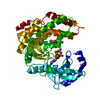
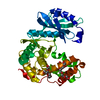
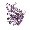
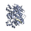
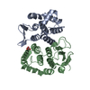
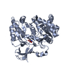
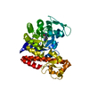
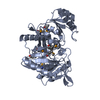
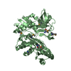
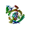
| Title | Amicoumacin kinase AmiN in complex with ATP | ||||||||||||
 Components Components | Phosphotransferase enzyme family protein, amicoumacin kinase | ||||||||||||
 Keywords Keywords | ANTIMICROBIAL PROTEIN / KINASE / AMICOUMACIN | ||||||||||||
| Function / homology |  Function and homology information Function and homology informationhomoserine kinase activity / L-threonine biosynthetic process / ATP binding / metal ion binding Similarity search - Function | ||||||||||||
| Biological species |  | ||||||||||||
| Method |  X-RAY DIFFRACTION / SYNCHROTRON / MOLECULAR REPLACEMENT / Resolution: 2 Å X-RAY DIFFRACTION / SYNCHROTRON / MOLECULAR REPLACEMENT / Resolution: 2 Å | ||||||||||||
 Authors Authors | Bourenkov, G.P. / Mokrushina, Y.A. / Terekhov, S.S. / Smirnov, I.V. / Gabibov, A.G. / Altman, S. | ||||||||||||
| Funding support |  Russian Federation, 3items Russian Federation, 3items
| ||||||||||||
 Citation Citation | Journal: Sci Adv / Year: 2020 Title: A kinase bioscavenger provides antibiotic resistance by extremely tight substrate binding. Authors: Terekhov, S.S. / Mokrushina, Y.A. / Nazarov, A.S. / Zlobin, A. / Zalevsky, A. / Bourenkov, G. / Golovin, A. / Belogurov Jr., A. / Osterman, I.A. / Kulikova, A.A. / Mitkevich, V.A. / Lou, H.J. ...Authors: Terekhov, S.S. / Mokrushina, Y.A. / Nazarov, A.S. / Zlobin, A. / Zalevsky, A. / Bourenkov, G. / Golovin, A. / Belogurov Jr., A. / Osterman, I.A. / Kulikova, A.A. / Mitkevich, V.A. / Lou, H.J. / Turk, B.E. / Wilmanns, M. / Smirnov, I.V. / Altman, S. / Gabibov, A.G. | ||||||||||||
| History |
|
- Structure visualization
| Structure viewer | Molecule: MolmilJmol/JSmol |
|---|
- Downloads & links
-Download
| PDBx/mmCIF format | 6sv5.cif.gz | 148.7 KB | Display | PDBx/mmCIF format |
|---|---|---|---|---|
| PDB format | pdb6sv5.ent.gz | 115.4 KB | Display | PDB format |
| PDBx/mmJSON format | 6sv5.json.gz | Tree view | PDBx/mmJSON format | |
| Others |  Other downloads Other downloads |
-Validation report
| Arichive directory | https://data.pdbj.org/pub/pdb/validation_reports/sv/6sv5ftp://data.pdbj.org/pub/pdb/validation_reports/sv/6sv5 | HTTPS FTP |
|---|
-Related structure data


-Links
 PDBj
PDBj
- Assembly
| Deposited unit | 
| ||||||||
|---|---|---|---|---|---|---|---|---|---|
| 1 |
| ||||||||
| Unit cell |
|
-Components
| #1: Protein | Mass: 39110.996 Da / Num. of mol.: 1 Source method: isolated from a genetically manipulated source Source: (gene. exp.) References: UniProt: A0A2T0D6W6, UniProt: A8FAR5*PLUS, EC: 2.7.1.230 |
|---|---|
| #2: Chemical | ChemComp-ATP /   Mass: 507.181 Da / Num. of mol.: 1 / Source method: obtained synthetically / Formula: C10H16N5O13P3 / Comment: ATP, energy-carrying molecule*YM Mass: 507.181 Da / Num. of mol.: 1 / Source method: obtained synthetically / Formula: C10H16N5O13P3 / Comment: ATP, energy-carrying molecule*YM |
| #3: Water | ChemComp-HOH /  Mass: 18.015 Da / Num. of mol.: 209 / Source method: isolated from a natural source / Formula: H2O Mass: 18.015 Da / Num. of mol.: 209 / Source method: isolated from a natural source / Formula: H2O |
| Has ligand of interest | N |
-Experimental details
-Experiment
| Experiment | Method: X-RAY DIFFRACTION / Number of used crystals: 1 |
|---|
- Sample preparation
| Crystal | Density Matthews: 3.11 Å3/Da / Density % sol: 60.48 % |
|---|---|
| Crystal grow | Temperature: 293 K / Method: vapor diffusion, hanging drop / pH: 8.5 Details: 100 mM Tris-HCl, pH 8.5 2.5-2.65 M ammonium sulfate 10 mM ATP |
-Data collection
| Diffraction | Mean temperature: 100 K / Serial crystal experiment: N |
|---|---|
| Diffraction source | Source: SYNCHROTRON / Site: PETRA III, EMBL c/o DESY  / Beamline: P13 (MX1) / Wavelength: 0.976 Å / Beamline: P13 (MX1) / Wavelength: 0.976 Å |
| Detector | Type: DECTRIS PILATUS 6M-F / Detector: PIXEL / Date: May 20, 2018 / Details: KB Mirrors |
| Radiation | Monochromator: Si 111 / Protocol: SINGLE WAVELENGTH / Monochromatic (M) / Laue (L): M / Scattering type: x-ray |
| Radiation wavelength | Wavelength: 0.976 Å / Relative weight: 1 |
| Reflection | Resolution: 2→70.5 Å / Num. obs: 34143 / % possible obs: 97.23 % / Redundancy: 6.9 % / Biso Wilson estimate: 25.4 Å2 / CC1/2: 0.999 / Rmerge(I) obs: 0.044 / Rrim(I) all: 0.048 / Net I/σ(I): 23.9 |
| Reflection shell | Resolution: 2→2.05 Å / Rmerge(I) obs: 0.213 / Mean I/σ(I) obs: 8.6 / Num. unique obs: 2366 / CC1/2: 0.987 / Rrim(I) all: 0.229 |
- Processing
| Software |
| ||||||||||||||||||||||||||||||||||||||||||||||||||||||||||||
|---|---|---|---|---|---|---|---|---|---|---|---|---|---|---|---|---|---|---|---|---|---|---|---|---|---|---|---|---|---|---|---|---|---|---|---|---|---|---|---|---|---|---|---|---|---|---|---|---|---|---|---|---|---|---|---|---|---|---|---|---|---|
| Refinement | Method to determine structure: MOLECULAR REPLACEMENT / Resolution: 2→70.48 Å / Cor.coef. Fo:Fc: 0.945 / Cor.coef. Fo:Fc free: 0.932 / SU B: 8.574 / SU ML: 0.121 / Cross valid method: THROUGHOUT / σ(F): 0 / ESU R: 0.167 / ESU R Free: 0.151 / Stereochemistry target values: MAXIMUM LIKELIHOOD Details: U VALUES : WITH TLS ADDED HYDROGENS HAVE BEEN ADDED IN THE RIDING POSITIONS
| ||||||||||||||||||||||||||||||||||||||||||||||||||||||||||||
| Solvent computation | Ion probe radii: 0.8 Å / Shrinkage radii: 0.8 Å / VDW probe radii: 1.2 Å / Solvent model: MASK | ||||||||||||||||||||||||||||||||||||||||||||||||||||||||||||
| Displacement parameters | Biso max: 123.57 Å2 / Biso mean: 32.7 Å2 / Biso min: 11.22 Å2
| ||||||||||||||||||||||||||||||||||||||||||||||||||||||||||||
| Refinement step | Cycle: final / Resolution: 2→70.48 Å
| ||||||||||||||||||||||||||||||||||||||||||||||||||||||||||||
| Refine LS restraints |
| ||||||||||||||||||||||||||||||||||||||||||||||||||||||||||||
| LS refinement shell | Resolution: 2→2.052 Å / Rfactor Rfree error: 0 / Total num. of bins used: 20
| ||||||||||||||||||||||||||||||||||||||||||||||||||||||||||||
| Refinement TLS params. | Method: refined / Origin x: 6.301 Å / Origin y: 16.774 Å / Origin z: 17.886 Å
|
