 Movie
Movie Controller


+ Open data

- Basic information
| Entry | Database: PDB / ID: 1vdf | ||||||
|---|---|---|---|---|---|---|---|
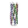
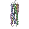
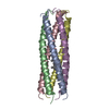
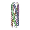
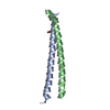
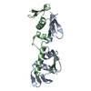
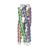
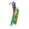
| Title | ASSEMBLY DOMAIN OF CARTILAGE OLIGOMERIC MATRIX PROTEIN | ||||||
 Components Components | CARTILAGE OLIGOMERIC MATRIX PROTEIN | ||||||
 Keywords Keywords | EXTRACELLULAR MATRIX PROTEIN / ASSEMBLY DOMAIN / CARTILAGE / OLIGOMERIC MATRIX PROTEIN / GLYCOPROTEIN | ||||||
| Function / homology |  Function and homology information Function and homology informationtendon development / negative regulation of hemostasis / cartilage homeostasis / Integrin cell surface interactions / ECM proteoglycans / vascular associated smooth muscle contraction / musculoskeletal movement / chondrocyte development / vascular associated smooth muscle cell development / BMP binding ...tendon development / negative regulation of hemostasis / cartilage homeostasis / Integrin cell surface interactions / ECM proteoglycans / vascular associated smooth muscle contraction / musculoskeletal movement / chondrocyte development / vascular associated smooth muscle cell development / BMP binding / bone growth / growth plate cartilage development / chondrocyte proliferation / endochondral bone growth / positive regulation of chondrocyte proliferation / regulation of bone mineralization / muscle cell development / vitamin D binding / bone morphogenesis / heparan sulfate proteoglycan binding / collagen fibril organization / proteoglycan binding / cartilage development / artery morphogenesis / skin development / extracellular matrix structural constituent / neuromuscular process / limb development / bone mineralization / fibronectin binding / protein secretion / BMP signaling pathway / response to unfolded protein / collagen binding / ossification / multicellular organism growth / skeletal system development / protein processing / protein homooligomerization / platelet aggregation / integrin binding / cellular senescence / blood coagulation / heparin binding / extracellular matrix / regulation of gene expression / protease binding / calcium ion binding / apoptotic process / negative regulation of apoptotic process / protein-containing complex / : / extracellular region Similarity search - Function | ||||||
| Biological species |  | ||||||
| Method |  X-RAY DIFFRACTION / MIRAS / Resolution: 2.05 Å X-RAY DIFFRACTION / MIRAS / Resolution: 2.05 Å | ||||||
 Authors Authors | Malashkevich, V.N. | ||||||
 Citation Citation | Journal: Science / Year: 1996 Title: The crystal structure of a five-stranded coiled coil in COMP: a prototype ion channel? Authors: Malashkevich, V.N. / Kammerer, R.A. / Efimov, V.P. / Schulthess, T. / Engel, J. #1: Journal: Proteins / Year: 1996Title: Crystallization and Preliminary Crystallographic Study of the Pentamerizing Domain from Cartilage Oligomeric Matrix Protein: A Five-Stranded Alpha-Helical Bundle Authors: Efimov, V.P. / Engel, J. / Malashkevich, V.N. | ||||||
| History |
|
- Structure visualization
| Structure viewer | Molecule: MolmilJmol/JSmol |
|---|
- Downloads & links
-Download
| PDBx/mmCIF format | 1vdf.cif.gz | 59.8 KB | Display | PDBx/mmCIF format |
|---|---|---|---|---|
| PDB format | pdb1vdf.ent.gz | 45.1 KB | Display | PDB format |
| PDBx/mmJSON format | 1vdf.json.gz | Tree view | PDBx/mmJSON format | |
| Others |  Other downloads Other downloads |
-Validation report
| Arichive directory | https://data.pdbj.org/pub/pdb/validation_reports/vd/1vdfftp://data.pdbj.org/pub/pdb/validation_reports/vd/1vdf | HTTPS FTP |
|---|
-Related structure data
| Similar structure data |
|---|


-Links
 PDBj
PDBj


- Assembly
| Deposited unit | 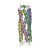
| ||||||||
|---|---|---|---|---|---|---|---|---|---|
| 1 |
| ||||||||
| Unit cell |
|
-Components
| #1: Protein/peptide | Mass: 5299.129 Da / Num. of mol.: 5 / Fragment: ASSEMBLY DOMAIN Source method: isolated from a genetically manipulated source Details: PENTAMERIC COILED-COIL / Source: (gene. exp.)  #2: Chemical | ChemComp-CL / |   Mass: 35.453 Da / Num. of mol.: 1 / Source method: obtained synthetically / Formula: Cl Mass: 35.453 Da / Num. of mol.: 1 / Source method: obtained synthetically / Formula: Cl#3: Water | ChemComp-HOH / |  Mass: 18.015 Da / Num. of mol.: 169 / Source method: isolated from a natural source / Formula: H2O Mass: 18.015 Da / Num. of mol.: 169 / Source method: isolated from a natural source / Formula: H2OHas protein modification | Y | |
|---|
-Experimental details
-Experiment
| Experiment | Method: X-RAY DIFFRACTION / Number of used crystals: 1 |
|---|
- Sample preparation
| Crystal | Density Matthews: 1.91 Å3/Da / Density % sol: 36 % / Description: MOLECULAR REPLACEMENT WAS NOT USED. | ||||||||||||||||||||||||||||||||||||||||||||||||||||||||||||
|---|---|---|---|---|---|---|---|---|---|---|---|---|---|---|---|---|---|---|---|---|---|---|---|---|---|---|---|---|---|---|---|---|---|---|---|---|---|---|---|---|---|---|---|---|---|---|---|---|---|---|---|---|---|---|---|---|---|---|---|---|---|
| Crystal grow | Method: vapor diffusion, hanging drop / pH: 6 Details: 17% PEG-1500, 50 MM NA-PI, PH 6.0, 0.5M NACL, HANGING DROP, vapor diffusion - hanging drop | ||||||||||||||||||||||||||||||||||||||||||||||||||||||||||||
| Crystal grow | *PLUS pH: 8.5 / Method: vapor diffusion, hanging dropDetails: Efimov, V.P., (1996) Proteins: Struct.,Funct., Genet., 24, 259. | ||||||||||||||||||||||||||||||||||||||||||||||||||||||||||||
| Components of the solutions | *PLUS
|
-Data collection
| Diffraction | Mean temperature: 279 K |
|---|---|
| Diffraction source | Source: ROTATING ANODE / Type: ELLIOTT / Wavelength: 1.5418 |
| Detector | Type: MARRESEARCH / Detector: IMAGE PLATE / Date: Dec 12, 1994 |
| Radiation | Monochromator: GRAPHITE(002) / Monochromatic (M) / Laue (L): M / Scattering type: x-ray |
| Radiation wavelength | Wavelength: 1.5418 Å / Relative weight: 1 |
| Reflection | Resolution: 2.05→25 Å / Num. obs: 12978 / % possible obs: 97 % / Observed criterion σ(I): 0 / Redundancy: 3.8 % / Biso Wilson estimate: 31.7 Å2 / Rmerge(I) obs: 0.04 / Rsym value: 0.04 / Net I/σ(I): 10.5 |
| Reflection shell | Resolution: 2.05→2.11 Å / Redundancy: 3.8 % / Mean I/σ(I) obs: 3.9 / Rsym value: 0.21 / % possible all: 81 |
| Reflection | *PLUS Num. measured all: 46638 |
- Processing
| Software |
| ||||||||||||||||||||||||||||||||||||||||||||||||||
|---|---|---|---|---|---|---|---|---|---|---|---|---|---|---|---|---|---|---|---|---|---|---|---|---|---|---|---|---|---|---|---|---|---|---|---|---|---|---|---|---|---|---|---|---|---|---|---|---|---|---|---|
| Refinement | Method to determine structure: MIRAS / Resolution: 2.05→8 Å / Isotropic thermal model: TNT / σ(F): 0 / Stereochemistry target values: ENGH & HUBER Details: MET 27 AND GLY 72 ARE DISORDERED IN ALL FIVE CHAINS OF THE CRYSTAL STRUCTURE. ALTHOUGH THEY WERE MODELED BASED ON THE EXISTING MAP, THEIR CONFORMATIONS ARE NOT SECURE. GLU 28 IN CHAIN D HAS ...Details: MET 27 AND GLY 72 ARE DISORDERED IN ALL FIVE CHAINS OF THE CRYSTAL STRUCTURE. ALTHOUGH THEY WERE MODELED BASED ON THE EXISTING MAP, THEIR CONFORMATIONS ARE NOT SECURE. GLU 28 IN CHAIN D HAS UNFAVORABLE PHI-PSI TORSION ANGLES. IT IS POORLY DEFINED IN THE ELECTRON DENSITY MAP.
| ||||||||||||||||||||||||||||||||||||||||||||||||||
| Solvent computation | Solvent model: TNT / Bsol: 171.3 Å2 / ksol: 0.826 e/Å3 | ||||||||||||||||||||||||||||||||||||||||||||||||||
| Refinement step | Cycle: LAST / Resolution: 2.05→8 Å
| ||||||||||||||||||||||||||||||||||||||||||||||||||
| Refine LS restraints |
| ||||||||||||||||||||||||||||||||||||||||||||||||||
| Software | *PLUS Name: TNT / Classification: refinement | ||||||||||||||||||||||||||||||||||||||||||||||||||
| Refinement | *PLUS Rfactor all: 0.176 | ||||||||||||||||||||||||||||||||||||||||||||||||||
| Solvent computation | *PLUS | ||||||||||||||||||||||||||||||||||||||||||||||||||
| Displacement parameters | *PLUS | ||||||||||||||||||||||||||||||||||||||||||||||||||
| Refine LS restraints | *PLUS
|
