 Movie
Movie Controller

[English] 日本語
 Yorodumi
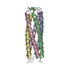
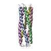
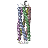
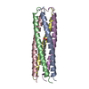
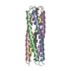
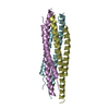
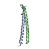
- PDB-1fbm: ASSEMBLY DOMAIN OF CARTILAGE OLIGOMERIC MATRIX PROTEIN IN COMPLEX... -
+ Open data

- Basic information
| Entry | Database: PDB / ID: 1fbm | ||||||
|---|---|---|---|---|---|---|---|
| Title | ASSEMBLY DOMAIN OF CARTILAGE OLIGOMERIC MATRIX PROTEIN IN COMPLEX WITH ALL-TRANS RETINOL | ||||||
 Components Components | PROTEIN (CARTILAGE OLIGOMERIC MATRIX PROTEIN) | ||||||
 Keywords Keywords | CELL ADHESION / EXTRACELLULAR MATRIX PROTEIN / ASSEMBLY DOMAIN / CARTILAGE / OLIGOMERIC MATRIX PROTEIN / GLYCOPROTEIN / RETINOL-COMPLEX | ||||||
| Function / homology |  Function and homology information Function and homology informationtendon development / negative regulation of hemostasis / cartilage homeostasis / Integrin cell surface interactions / ECM proteoglycans / vascular associated smooth muscle contraction / musculoskeletal movement / chondrocyte development / vascular associated smooth muscle cell development / BMP binding ...tendon development / negative regulation of hemostasis / cartilage homeostasis / Integrin cell surface interactions / ECM proteoglycans / vascular associated smooth muscle contraction / musculoskeletal movement / chondrocyte development / vascular associated smooth muscle cell development / BMP binding / bone growth / growth plate cartilage development / chondrocyte proliferation / endochondral bone growth / positive regulation of chondrocyte proliferation / regulation of bone mineralization / muscle cell development / vitamin D binding / bone morphogenesis / heparan sulfate proteoglycan binding / collagen fibril organization / proteoglycan binding / cartilage development / artery morphogenesis / skin development / extracellular matrix structural constituent / neuromuscular process / limb development / bone mineralization / fibronectin binding / protein secretion / BMP signaling pathway / response to unfolded protein / collagen binding / ossification / multicellular organism growth / skeletal system development / protein processing / protein homooligomerization / platelet aggregation / integrin binding / cellular senescence / blood coagulation / heparin binding / extracellular matrix / regulation of gene expression / protease binding / calcium ion binding / apoptotic process / negative regulation of apoptotic process / protein-containing complex / : / extracellular region Similarity search - Function | ||||||
| Biological species |  | ||||||
| Method |  X-RAY DIFFRACTION / Resolution: 2.7 Å X-RAY DIFFRACTION / Resolution: 2.7 Å | ||||||
 Authors Authors | Guo, Y. / Bozic, D. / Malashkevich, V.N. / Kammerer, R.A. / Schulthess, T. | ||||||
 Citation Citation | Journal: EMBO J. / Year: 1998 Title: All-trans retinol, vitamin D and other hydrophobic compounds bind in the axial pore of the five-stranded coiled-coil domain of cartilage oligomeric matrix protein. Authors: Guo, Y. / Bozic, D. / Malashkevich, V.N. / Kammerer, R.A. / Schulthess, T. #1: Journal: Proteins / Year: 1996Title: Crystallization and Preliminary Crystallographic Study of the Pentamerizing Domain from Cartilage Oligomeric Matrix Protein: a Five-stranded Alpha-helical Bundle. Authors: Efimov, V.P. / Engel, J. / Malashkevich, V.N. #2: Journal: Science / Year: 1996Title: The Crystal Structure of a Five-stranded Coiled Coil in COMP: a Prototype Ion Channel? Authors: Malashkevich, V.N. / Kammerer, R.A. / Efimov, V.P. / Schulthess, T. / Engel, J. | ||||||
| History |
|
- Structure visualization
| Structure viewer | Molecule: MolmilJmol/JSmol |
|---|
- Downloads & links
-Download
| PDBx/mmCIF format | 1fbm.cif.gz | 59.5 KB | Display | PDBx/mmCIF format |
|---|---|---|---|---|
| PDB format | pdb1fbm.ent.gz | 45.3 KB | Display | PDB format |
| PDBx/mmJSON format | 1fbm.json.gz | Tree view | PDBx/mmJSON format | |
| Others |  Other downloads Other downloads |
-Validation report
| Arichive directory | https://data.pdbj.org/pub/pdb/validation_reports/fb/1fbmftp://data.pdbj.org/pub/pdb/validation_reports/fb/1fbm | HTTPS FTP |
|---|
-Related structure data
| Related structure data | |
|---|---|
| Similar structure data |


-Links
 PDBj
PDBj


- Assembly
| Deposited unit | 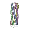
| ||||||||
|---|---|---|---|---|---|---|---|---|---|
| 1 |
| ||||||||
| Unit cell |
|
-Components
| #1: Protein/peptide | Mass: 5299.129 Da / Num. of mol.: 5 / Fragment: ASSEMBLY DOMAIN Source method: isolated from a genetically manipulated source Details: PENTAMERIC COILED-COIL / Source: (gene. exp.)  #2: Chemical | ChemComp-RTL / |   Mass: 286.452 Da / Num. of mol.: 1 / Source method: obtained synthetically / Formula: C20H30O Mass: 286.452 Da / Num. of mol.: 1 / Source method: obtained synthetically / Formula: C20H30O#3: Water | ChemComp-HOH / |  Mass: 18.015 Da / Num. of mol.: 152 / Source method: isolated from a natural source / Formula: H2O Mass: 18.015 Da / Num. of mol.: 152 / Source method: isolated from a natural source / Formula: H2OHas protein modification | Y | |
|---|
-Experimental details
-Experiment
| Experiment | Method: X-RAY DIFFRACTION / Number of used crystals: 1 |
|---|
- Sample preparation
| Crystal | Density Matthews: 1.92 Å3/Da / Density % sol: 35.83 % | ||||||||||||||||||||||||||||||
|---|---|---|---|---|---|---|---|---|---|---|---|---|---|---|---|---|---|---|---|---|---|---|---|---|---|---|---|---|---|---|---|
| Crystal grow | Temperature: 293 K / Method: vapor diffusion, hanging drop / pH: 6 Details: COMPcc (16 mg/ml) in 20 mM sodium phosphate, 200 mM NaCl (pH 7.5) precipitant: 7-8% PEG 8000, 50 mM sodium cacodylate (pH 6.0), 50 mM sodium acetate, 50 mM calcium acetate , VAPOR DIFFUSION, ...Details: COMPcc (16 mg/ml) in 20 mM sodium phosphate, 200 mM NaCl (pH 7.5) precipitant: 7-8% PEG 8000, 50 mM sodium cacodylate (pH 6.0), 50 mM sodium acetate, 50 mM calcium acetate , VAPOR DIFFUSION, HANGING DROP, temperature 20.0K | ||||||||||||||||||||||||||||||
| Crystal grow | *PLUS | ||||||||||||||||||||||||||||||
| Components of the solutions | *PLUS
|
-Data collection
| Diffraction | Mean temperature: 298 K |
|---|---|
| Diffraction source | Source: ROTATING ANODE / Type: ELLIOTT GX-20 / Wavelength: 1.5418 / Wavelength: 1.5418 Å |
| Detector | Type: MARRESEARCH / Detector: IMAGE PLATE / Date: Dec 1, 1997 |
| Radiation | Protocol: SINGLE WAVELENGTH / Monochromatic (M) / Laue (L): M / Scattering type: x-ray |
| Radiation wavelength | Wavelength: 1.5418 Å / Relative weight: 1 |
| Reflection | Resolution: 2.7→3 Å / Num. all: 5464 / Num. obs: 5464 / % possible obs: 86 % / Redundancy: 3.71 % / Biso Wilson estimate: 32.2 Å2 / Rmerge(I) obs: 0.132 |
| Reflection shell | Resolution: 2.7→3 Å / Num. unique all: 5464 / % possible all: 87.6 |
| Reflection | *PLUS Num. measured all: 20313 |
| Reflection shell | *PLUS % possible obs: 87.6 % |
- Processing
| Software |
| |||||||||||||||||||||
|---|---|---|---|---|---|---|---|---|---|---|---|---|---|---|---|---|---|---|---|---|---|---|
| Refinement | Resolution: 2.7→3 Å / σ(F): 0 / Stereochemistry target values: Engh & Huber
| |||||||||||||||||||||
| Refinement step | Cycle: LAST / Resolution: 2.7→3 Å
| |||||||||||||||||||||
| Refine LS restraints |
| |||||||||||||||||||||
| Software | *PLUS Name: X-PLOR / Version: 3.1 / Classification: refinement | |||||||||||||||||||||
| Refine LS restraints | *PLUS
|
