 Movie
Movie Controller

[English] 日本語
 Yorodumi
- PDB-1vaj: Crystal Structure of Uncharacterized Protein PH0010 From Pyrococc... -
+ Open data

- Basic information
| Entry | Database: PDB / ID: 1vaj | ||||||
|---|---|---|---|---|---|---|---|
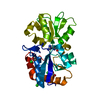
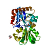
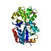
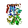
| Title | Crystal Structure of Uncharacterized Protein PH0010 From Pyrococcus horikoshii | ||||||
 Components Components | Hypothetical protein PH0010 | ||||||
 Keywords Keywords | STRUCTURAL GENOMICS / UNKNOWN FUNCTION / alpha + beta fold | ||||||
| Function / homology |  Function and homology information Function and homology informationHypothetical protein ph0010; domain 2 / Hypothetical protein ph0010; domain 1 / AMMECR1 domain / Uncharacterised protein family MJ0810 / AMMECR1 / AMMECR1, N-terminal / AmmeMemoRadiSam system protein A / AMMECR1 domain superfamily / AMMECR1 / AMMECR1 domain profile. ...Hypothetical protein ph0010; domain 2 / Hypothetical protein ph0010; domain 1 / AMMECR1 domain / Uncharacterised protein family MJ0810 / AMMECR1 / AMMECR1, N-terminal / AmmeMemoRadiSam system protein A / AMMECR1 domain superfamily / AMMECR1 / AMMECR1 domain profile. / Glycoprotein, Type 4 Pilin / Dna Ligase; domain 1 / 2-Layer Sandwich / Alpha Beta Similarity search - Domain/homology | ||||||
| Biological species |   Pyrococcus horikoshii (archaea) Pyrococcus horikoshii (archaea) | ||||||
| Method |  X-RAY DIFFRACTION / SYNCHROTRON / MAD / Resolution: 1.82 Å X-RAY DIFFRACTION / SYNCHROTRON / MAD / Resolution: 1.82 Å | ||||||
 Authors Authors | Tajika, Y. / Sakai, N. / Tamura, T. / Yao, M. / Watanabe, N. / Tanaka, I. | ||||||
 Citation Citation | Journal: Proteins / Year: 2005 Title: Crystal structure of PH0010 from Pyrococcus horikoshii, which is highly homologous to human AMMECR 1C-terminal region Authors: Tajika, Y. / Sakai, N. / Tamura, T. / Yao, M. / Watanabe, N. / Tanaka, I. | ||||||
| History |
|
- Structure visualization
| Structure viewer | Molecule: MolmilJmol/JSmol |
|---|
- Downloads & links
-Download
| PDBx/mmCIF format | 1vaj.cif.gz | 57 KB | Display | PDBx/mmCIF format |
|---|---|---|---|---|
| PDB format | pdb1vaj.ent.gz | 40.9 KB | Display | PDB format |
| PDBx/mmJSON format | 1vaj.json.gz | Tree view | PDBx/mmJSON format | |
| Others |  Other downloads Other downloads |
-Validation report
| Summary document | 1vaj_validation.pdf.gz | 369 KB | Display | wwPDB validaton report |
|---|---|---|---|---|
| Full document | 1vaj_full_validation.pdf.gz | 370.9 KB | Display | |
| Data in XML | 1vaj_validation.xml.gz | 5.6 KB | Display | |
| Data in CIF | 1vaj_validation.cif.gz | 8.7 KB | Display | |
| Arichive directory | https://data.pdbj.org/pub/pdb/validation_reports/va/1vajftp://data.pdbj.org/pub/pdb/validation_reports/va/1vaj | HTTPS FTP |
-Related structure data
| Similar structure data |
|---|


-Links
 PDBj
PDBj- Assembly
| Deposited unit | 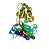
| ||||||||
|---|---|---|---|---|---|---|---|---|---|
| 1 |
| ||||||||
| Unit cell |
|
-Components
| #1: Protein | Mass: 24862.168 Da / Num. of mol.: 1 Source method: isolated from a genetically manipulated source Source: (gene. exp.) Pyrococcus horikoshii (archaea) / Gene: PH0010 / Plasmid: pET-26b(+) / Production host:  Strain (production host): BL21-codonplus(DE3)-RIL (Stratagene) References: UniProt: O57770 |
|---|---|
| #2: Water | ChemComp-HOH /  Mass: 18.015 Da / Num. of mol.: 165 / Source method: isolated from a natural source / Formula: H2O Mass: 18.015 Da / Num. of mol.: 165 / Source method: isolated from a natural source / Formula: H2O |
| Has protein modification | Y |
-Experimental details
-Experiment
| Experiment | Method: X-RAY DIFFRACTION / Number of used crystals: 1 |
|---|
- Sample preparation
| Crystal | Density Matthews: 2.46 Å3/Da / Density % sol: 50 % |
|---|---|
| Crystal grow | Temperature: 293 K / Method: vapor diffusion, hanging drop / pH: 7.9 Details: PEG 8000, magnesium chloride, pH 7.9, VAPOR DIFFUSION, HANGING DROP, temperature 293K |
-Data collection
| Diffraction | Mean temperature: 100 K | ||||||||||||
|---|---|---|---|---|---|---|---|---|---|---|---|---|---|
| Diffraction source | Source: SYNCHROTRON / Site: Photon Factory  / Beamline: AR-NW12A / Wavelength: 0.9742, 0.9785, 0.9600 / Beamline: AR-NW12A / Wavelength: 0.9742, 0.9785, 0.9600 | ||||||||||||
| Detector | Detector: CCD / Date: Dec 21, 2003 | ||||||||||||
| Radiation | Protocol: MAD / Monochromatic (M) / Laue (L): M / Scattering type: x-ray | ||||||||||||
| Radiation wavelength |
| ||||||||||||
| Reflection | Resolution: 1.82→38.63 Å / Num. obs: 19449 / % possible obs: 95.7 % / Observed criterion σ(I): -3 / Redundancy: 5.4 % / Biso Wilson estimate: 26.5 Å2 / Rmerge(I) obs: 0.071 / Net I/σ(I): 10.6 | ||||||||||||
| Reflection shell | Resolution: 1.82→1.89 Å / Redundancy: 3.8 % / Rmerge(I) obs: 0.336 / % possible all: 77.2 |
- Processing
| Software |
| |||||||||||||||||||||||||||
|---|---|---|---|---|---|---|---|---|---|---|---|---|---|---|---|---|---|---|---|---|---|---|---|---|---|---|---|---|
| Refinement | Method to determine structure: MAD / Resolution: 1.82→10 Å / Cross valid method: THROUGHOUT / σ(F): 0 / Stereochemistry target values: Engh & Huber
| |||||||||||||||||||||||||||
| Displacement parameters | Biso mean: 20.24 Å2
| |||||||||||||||||||||||||||
| Refine analyze |
| |||||||||||||||||||||||||||
| Refinement step | Cycle: LAST / Resolution: 1.82→10 Å
| |||||||||||||||||||||||||||
| Refine LS restraints |
| |||||||||||||||||||||||||||
| LS refinement shell | Resolution: 1.82→1.88 Å / Total num. of bins used: 10
| |||||||||||||||||||||||||||
| Xplor file | Serial no: 1 / Param file: protein_rep.param / Topol file: protein.top |
