 Movie
Movie Controller

[English] 日本語
 Yorodumi
- PDB-1v7u: Crystal structure of Undecaprenyl Pyrophosphate Synthase with far... -
+ Open data

- Basic information
| Entry | Database: PDB / ID: 1v7u | ||||||
|---|---|---|---|---|---|---|---|
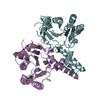
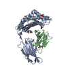
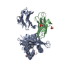
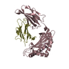
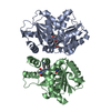
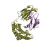
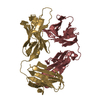
| Title | Crystal structure of Undecaprenyl Pyrophosphate Synthase with farnesyl pyrophosphate | ||||||
 Components Components | Undecaprenyl pyrophosphate synthetase | ||||||
 Keywords Keywords | TRANSFERASE / prenyltransferase / farnesyl pyrophosphate / isopentenyl pyrophosphate / substrate binding | ||||||
| Function / homology |  Function and homology information Function and homology informationGram-negative-bacterium-type cell wall biogenesis / ditrans,polycis-undecaprenyl-diphosphate synthase [(2E,6E)-farnesyl-diphosphate specific] / ditrans,polycis-undecaprenyl-diphosphate synthase [(2E,6E)-farnesyl-diphosphate specific] activity / polyprenol biosynthetic process / small molecule binding / peptidoglycan biosynthetic process / cell wall organization / regulation of cell shape / cell division / magnesium ion binding ...Gram-negative-bacterium-type cell wall biogenesis / ditrans,polycis-undecaprenyl-diphosphate synthase [(2E,6E)-farnesyl-diphosphate specific] / ditrans,polycis-undecaprenyl-diphosphate synthase [(2E,6E)-farnesyl-diphosphate specific] activity / polyprenol biosynthetic process / small molecule binding / peptidoglycan biosynthetic process / cell wall organization / regulation of cell shape / cell division / magnesium ion binding / protein homodimerization activity / cytosol / cytoplasm Similarity search - Function | ||||||
| Biological species |  | ||||||
| Method |  X-RAY DIFFRACTION / AB INITIO PHASING / Resolution: 2.35 Å X-RAY DIFFRACTION / AB INITIO PHASING / Resolution: 2.35 Å | ||||||
 Authors Authors | Chang, S.-Y. / Ko, T.-P. / Chen, A.P.-C. / Wang, A.H.-J. / Liang, P.-H. | ||||||
 Citation Citation | Journal: Protein Sci. / Year: 2004 Title: Substrate binding mode and reaction mechanism of undecaprenyl pyrophosphate synthase deduced from crystallographic studies Authors: Chang, S.-Y. / Ko, T.-P. / Chen, A.P.-C. / Wang, A.H.-J. / Liang, P.-H. | ||||||
| History |
|
- Structure visualization
| Structure viewer | Molecule: MolmilJmol/JSmol |
|---|
- Downloads & links
-Download
| PDBx/mmCIF format | 1v7u.cif.gz | 111.9 KB | Display | PDBx/mmCIF format |
|---|---|---|---|---|
| PDB format | pdb1v7u.ent.gz | 86.4 KB | Display | PDB format |
| PDBx/mmJSON format | 1v7u.json.gz | Tree view | PDBx/mmJSON format | |
| Others |  Other downloads Other downloads |
-Validation report
| Arichive directory | https://data.pdbj.org/pub/pdb/validation_reports/v7/1v7uftp://data.pdbj.org/pub/pdb/validation_reports/v7/1v7u | HTTPS FTP |
|---|
-Related structure data
| Related structure data | |
|---|---|
| Similar structure data |


-Links
 PDBj
PDBj- Assembly
| Deposited unit | 
| ||||||||
|---|---|---|---|---|---|---|---|---|---|
| 1 |
| ||||||||
| Unit cell |
|
-Components
| #1: Protein | Mass: 28481.127 Da / Num. of mol.: 2 Source method: isolated from a genetically manipulated source Source: (gene. exp.) References: UniProt: P60472, ditrans,polycis-undecaprenyl-diphosphate synthase [(2E,6E)-farnesyl-diphosphate specific] #2: Chemical |   Mass: 382.326 Da / Num. of mol.: 3 / Source method: obtained synthetically / Formula: C15H28O7P2 Mass: 382.326 Da / Num. of mol.: 3 / Source method: obtained synthetically / Formula: C15H28O7P2#3: Water | ChemComp-HOH / |  Mass: 18.015 Da / Num. of mol.: 456 / Source method: isolated from a natural source / Formula: H2O Mass: 18.015 Da / Num. of mol.: 456 / Source method: isolated from a natural source / Formula: H2O |
|---|
-Experimental details
-Experiment
| Experiment | Method: X-RAY DIFFRACTION / Number of used crystals: 1 |
|---|
- Sample preparation
| Crystal | Density Matthews: 2.07 Å3/Da / Density % sol: 40.19 % | ||||||||||||||||||||||||||||||||||||||||||
|---|---|---|---|---|---|---|---|---|---|---|---|---|---|---|---|---|---|---|---|---|---|---|---|---|---|---|---|---|---|---|---|---|---|---|---|---|---|---|---|---|---|---|---|
| Crystal grow | Temperature: 298 K / Method: vapor diffusion, hanging drop / pH: 7 Details: 25% ethylene glycol, 0.05% triton, 0.5mM MgCl2, pH 7.0, VAPOR DIFFUSION, HANGING DROP, temperature 298K | ||||||||||||||||||||||||||||||||||||||||||
| Crystal grow | *PLUS Temperature: 25 ℃ / pH: 7.5 / Method: vapor diffusion, hanging drop | ||||||||||||||||||||||||||||||||||||||||||
| Components of the solutions | *PLUS
|
-Data collection
| Diffraction | Mean temperature: 123 K |
|---|---|
| Diffraction source | Source: ROTATING ANODE / Type: RIGAKU / Wavelength: 1.5 Å |
| Detector | Type: RIGAKU RAXIS IV / Detector: IMAGE PLATE |
| Radiation | Protocol: SINGLE WAVELENGTH / Monochromatic (M) / Laue (L): M / Scattering type: x-ray |
| Radiation wavelength | Wavelength: 1.5 Å / Relative weight: 1 |
| Reflection | Resolution: 2.35→10 Å / Num. all: 20047 / Num. obs: 19508 / % possible obs: 97.1 % / Observed criterion σ(I): 1 / Redundancy: 4.65 % / Rmerge(I) obs: 0.129 / Net I/σ(I): 11 |
| Reflection shell | Resolution: 2.35→2.43 Å / Redundancy: 4.65 % / Rmerge(I) obs: 0.129 / Mean I/σ(I) obs: 11 / Num. unique all: 19508 / % possible all: 97.1 |
| Reflection | *PLUS Num. measured all: 95659 |
| Reflection shell | *PLUS % possible obs: 95.1 % / Num. unique obs: 1882 / Num. measured obs: 9202 / Rmerge(I) obs: 0.363 / Mean I/σ(I) obs: 4.1 |
- Processing
| Software |
| |||||||||||||||||||||||||
|---|---|---|---|---|---|---|---|---|---|---|---|---|---|---|---|---|---|---|---|---|---|---|---|---|---|---|
| Refinement | Method to determine structure: AB INITIO PHASING / Resolution: 2.35→10 Å / σ(F): 0 / Stereochemistry target values: Engh & Huber
| |||||||||||||||||||||||||
| Refine analyze |
| |||||||||||||||||||||||||
| Refinement step | Cycle: LAST / Resolution: 2.35→10 Å
| |||||||||||||||||||||||||
| Refine LS restraints |
| |||||||||||||||||||||||||
| LS refinement shell | Resolution: 2.35→2.43 Å
| |||||||||||||||||||||||||
| Refinement | *PLUS % reflection Rfree: 5 % | |||||||||||||||||||||||||
| Solvent computation | *PLUS | |||||||||||||||||||||||||
| Displacement parameters | *PLUS | |||||||||||||||||||||||||
| LS refinement shell | *PLUS Num. reflection obs: 1786 |
