 Movie
Movie Controller


+ Open data

- Basic information
| Entry | Database: PDB / ID: 1uxa | |||||||||
|---|---|---|---|---|---|---|---|---|---|---|
| Title | ADENOVIRUS AD37 FIBRE HEAD in complex with sialyl-lactose | |||||||||
 Components Components | FIBER PROTEIN | |||||||||
 Keywords Keywords | VIRAL PROTEIN / ADENOVIRUS / AD37 / FIBRE / RECEPTOR / SIALIC ACID / NEURAMINIC ACID / CD46 / DAF / CONJUNCTIVITIS | |||||||||
| Function / homology |  Function and homology information Function and homology informationadhesion receptor-mediated virion attachment to host cell / viral capsid / cell adhesion / symbiont entry into host cell / host cell nucleus / metal ion binding Similarity search - Function | |||||||||
| Biological species |  HUMAN ADENOVIRUS TYPE 37 HUMAN ADENOVIRUS TYPE 37 | |||||||||
| Method |  X-RAY DIFFRACTION / SYNCHROTRON / MOLECULAR REPLACEMENT / Resolution: 1.5 Å X-RAY DIFFRACTION / SYNCHROTRON / MOLECULAR REPLACEMENT / Resolution: 1.5 Å | |||||||||
 Authors Authors | Burmeister, W.P. / Guilligay, D. / Cusack, S. / Wadell, G. / Arnberg, N. | |||||||||
 Citation Citation | Journal: J.Virol. / Year: 2004 Title: Crystal Structure of Species D Adenovirus Fiber Knobs and Their Sialic Acid Binding Sites Authors: Burmeister, W.P. / Guilligay, D. / Cusack, S. / Wadell, G. / Arnberg, N. | |||||||||
| History |
|
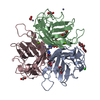
- Structure visualization
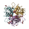
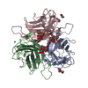
| Structure viewer | Molecule: MolmilJmol/JSmol |
|---|
- Downloads & links
-Download
| PDBx/mmCIF format | 1uxa.cif.gz | 135.4 KB | Display | PDBx/mmCIF format |
|---|---|---|---|---|
| PDB format | pdb1uxa.ent.gz | 105.3 KB | Display | PDB format |
| PDBx/mmJSON format | 1uxa.json.gz | Tree view | PDBx/mmJSON format | |
| Others |  Other downloads Other downloads |
-Validation report
| Summary document | 1uxa_validation.pdf.gz | 1.5 MB | Display | wwPDB validaton report |
|---|---|---|---|---|
| Full document | 1uxa_full_validation.pdf.gz | 1.5 MB | Display | |
| Data in XML | 1uxa_validation.xml.gz | 28.5 KB | Display | |
| Data in CIF | 1uxa_validation.cif.gz | 41.9 KB | Display | |
| Arichive directory | https://data.pdbj.org/pub/pdb/validation_reports/ux/1uxaftp://data.pdbj.org/pub/pdb/validation_reports/ux/1uxa | HTTPS FTP |
-Related structure data
| Related structure data |  1uxbC  1uxeC 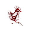 1qhvS C: citing same article ( S: Starting model for refinement |
|---|---|
| Similar structure data |


-Links
 PDBj
PDBj
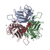
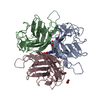
- Assembly
| Deposited unit | 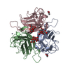
| ||||||||||||
|---|---|---|---|---|---|---|---|---|---|---|---|---|---|
| 1 |
| ||||||||||||
| Unit cell |
| ||||||||||||
| Noncrystallographic symmetry (NCS) | NCS oper:
|
-Components
-Protein , 1 types, 3 molecules ABC
| #1: Protein | Mass: 21716.535 Da / Num. of mol.: 3 / Fragment: HEAD DOMAIN, RESIDUES 172-365 / Mutation: YES Source method: isolated from a genetically manipulated source Details: ONLY THE KNOB DOMAIN, AFTER TEV CLEAVAGE OF A HIS-TAGGED PROTEIN CONSTRUCT Source: (gene. exp.) HUMAN ADENOVIRUS TYPE 37 / Strain: P / Plasmid: PPROEX HTB / Production host:  |
|---|
-Sugars , 2 types, 3 molecules
| #2: Polysaccharide | Source method: isolated from a genetically manipulated source #3: Polysaccharide | N-acetyl-alpha-neuraminic acid-(2-6)-beta-D-galactopyranose | Source method: isolated from a genetically manipulated source |
|---|
-Non-polymers , 3 types, 533 molecules 


| #4: Chemical |  Mass: 65.409 Da / Num. of mol.: 3 / Source method: obtained synthetically / Formula: Zn Mass: 65.409 Da / Num. of mol.: 3 / Source method: obtained synthetically / Formula: Zn#5: Chemical |  Mass: 59.044 Da / Num. of mol.: 2 / Source method: obtained synthetically / Formula: C2H3O2 Mass: 59.044 Da / Num. of mol.: 2 / Source method: obtained synthetically / Formula: C2H3O2#6: Water | ChemComp-HOH / | Mass: 18.015 Da / Num. of mol.: 528 / Source method: isolated from a natural source / Formula: H2O |
|---|
-Details
| Compound details | ENGINEERED MUTATION IN CHAINS A, B, C, TYR 173 TO ALA 173 ENGINEERED MUTATION IN CHAINS A, B, C, ...ENGINEERED |
|---|---|
| Sequence details | THE FIRST FIVE RESIDUES HAVE BEEN CHANGED IN ORDER TO INTRODUCE THE TEV CLEAVAGE SITE |
-Experimental details
-Experiment
| Experiment | Method: X-RAY DIFFRACTION / Number of used crystals: 1 |
|---|
- Sample preparation
| Crystal | Density Matthews: 2.43 Å3/Da / Density % sol: 49 % |
|---|---|
| Crystal grow | pH: 7.5 Details: RESERVOIR: 24 % PEG8000, 50 MM ZINC ACETATE, 100 MM HEPES, PROTEIN: 30 MM TRIS-HCL, PH 7.5, 150 MM NACL |
-Data collection
| Diffraction | Mean temperature: 100 K |
|---|---|
| Diffraction source | Source: SYNCHROTRON / Site: ESRF  / Beamline: ID29 / Wavelength: 0.979 / Beamline: ID29 / Wavelength: 0.979 |
| Detector | Type: ADSC CCD / Detector: CCD / Date: Apr 9, 2002 / Details: TOROIDAL MIRROR |
| Radiation | Monochromator: SI(111) / Protocol: SINGLE WAVELENGTH / Monochromatic (M) / Laue (L): M / Scattering type: x-ray |
| Radiation wavelength | Wavelength: 0.979 Å / Relative weight: 1 |
| Reflection | Resolution: 1.5→45.18 Å / Num. obs: 87661 / % possible obs: 92.3 % / Redundancy: 3.33 % / Biso Wilson estimate: 18.9 Å2 / Rmerge(I) obs: 0.073 / Net I/σ(I): 5.059 |
| Reflection shell | Resolution: 1.5→1.58 Å / Redundancy: 2.21 % / Rmerge(I) obs: 0.25 / Mean I/σ(I) obs: 2.21 / % possible all: 66.6 |
- Processing
| Software |
| ||||||||||||||||||||||||||||||||||||||||||||||||||||||||||||||||||||||||||||||||
|---|---|---|---|---|---|---|---|---|---|---|---|---|---|---|---|---|---|---|---|---|---|---|---|---|---|---|---|---|---|---|---|---|---|---|---|---|---|---|---|---|---|---|---|---|---|---|---|---|---|---|---|---|---|---|---|---|---|---|---|---|---|---|---|---|---|---|---|---|---|---|---|---|---|---|---|---|---|---|---|---|---|
| Refinement | Method to determine structure: MOLECULAR REPLACEMENT Starting model: PDB ENTRY 1QHV Resolution: 1.5→45.18 Å / Rfactor Rfree error: 0.003 / Isotropic thermal model: RESTRAINED / Cross valid method: THROUGHOUT / σ(F): 0 Details: THE ELECTRON DENSITY THAT IS ASSOCIATED WITH THE ACETATE IONS IS POORLY DEFINED
| ||||||||||||||||||||||||||||||||||||||||||||||||||||||||||||||||||||||||||||||||
| Solvent computation | Solvent model: FLAT MODEL / Bsol: 44.02 Å2 / ksol: 0.393 e/Å3 | ||||||||||||||||||||||||||||||||||||||||||||||||||||||||||||||||||||||||||||||||
| Displacement parameters | Biso mean: 22.81 Å2
| ||||||||||||||||||||||||||||||||||||||||||||||||||||||||||||||||||||||||||||||||
| Refine analyze |
| ||||||||||||||||||||||||||||||||||||||||||||||||||||||||||||||||||||||||||||||||
| Refinement step | Cycle: LAST / Resolution: 1.5→45.18 Å
| ||||||||||||||||||||||||||||||||||||||||||||||||||||||||||||||||||||||||||||||||
| Refine LS restraints |
| ||||||||||||||||||||||||||||||||||||||||||||||||||||||||||||||||||||||||||||||||
| LS refinement shell | Resolution: 1.5→1.55 Å / Rfactor Rfree error: 0.02 / Total num. of bins used: 10
| ||||||||||||||||||||||||||||||||||||||||||||||||||||||||||||||||||||||||||||||||
| Xplor file |
|
