[English] 日本語
Yorodumi
- PDB-1uxe: ADENOVIRUS AD37 FIBRE HEAD -

+
Open data


ID or keywords:

Loading...

-
Basic information

Entry
Database: PDB / ID: 1uxe
TitleADENOVIRUS AD37 FIBRE HEAD
ComponentsFIBER PROTEIN
KeywordsVIRAL PROTEIN / ADENOVIRUS / AD37 / FIBRE / RECEPTOR / SIALIC ACID / NEURAMINIC ACID / CD46 / DAF / CONJUNCTIVITIS
Function / homology
Function and homology information


adhesion receptor-mediated virion attachment to host cell / viral capsid / cell adhesion / symbiont entry into host cell / host cell nucleus / metal ion binding
Similarity search - Function
Adenovirus pIV-related, attachment domain / Adenovirus Type 5 Fiber Protein (Receptor Binding Domain) / Adenoviral fibre protein, knob / Adenoviral fibre protein (knob domain) / Adenoviral fibre protein, repeat/shaft region / Adenoviral fibre protein (repeat/shaft region) / Adenovirus fibre protein / Attachment protein shaft domain superfamily / Adenovirus pIV-like, attachment domain / Sandwich / Mainly Beta
Similarity search - Domain/homology
Biological speciesHUMAN ADENOVIRUS TYPE 37
MethodX-RAY DIFFRACTION / SYNCHROTRON / MOLECULAR REPLACEMENT / Resolution: 2 Å
AuthorsBurmeister, W.P. / Guilligay, D. / Cusack, S. / Wadell, G. / Arnberg, N.
CitationJournal: J.Virol. / Year: 2004
Title: Crystal Structure of Species D Adenovirus Fiber Knobs and Their Sialic Acid Binding Sites
Authors: Burmeister, W.P. / Guilligay, D. / Cusack, S. / Wadell, G. / Arnberg, N.
History
DepositionFeb 24, 2004Deposition site: PDBE / Processing site: PDBE
Revision 1.0Jul 2, 2004Provider: repository / Type: Initial release
Revision 1.1May 7, 2011Group: Version format compliance
Revision 1.2Jul 13, 2011Group: Version format compliance
Revision 1.3Dec 13, 2023Group: Data collection / Database references ...Data collection / Database references / Derived calculations / Other / Refinement description
Category: chem_comp_atom / chem_comp_bond ...chem_comp_atom / chem_comp_bond / database_2 / pdbx_database_status / pdbx_initial_refinement_model / pdbx_struct_conn_angle / struct_conn
Item: _database_2.pdbx_DOI / _database_2.pdbx_database_accession ..._database_2.pdbx_DOI / _database_2.pdbx_database_accession / _pdbx_database_status.status_code_sf / _pdbx_struct_conn_angle.ptnr1_auth_comp_id / _pdbx_struct_conn_angle.ptnr1_auth_seq_id / _pdbx_struct_conn_angle.ptnr1_label_asym_id / _pdbx_struct_conn_angle.ptnr1_label_atom_id / _pdbx_struct_conn_angle.ptnr1_label_comp_id / _pdbx_struct_conn_angle.ptnr1_label_seq_id / _pdbx_struct_conn_angle.ptnr3_auth_comp_id / _pdbx_struct_conn_angle.ptnr3_auth_seq_id / _pdbx_struct_conn_angle.ptnr3_label_asym_id / _pdbx_struct_conn_angle.ptnr3_label_atom_id / _pdbx_struct_conn_angle.ptnr3_label_comp_id / _pdbx_struct_conn_angle.ptnr3_label_seq_id / _pdbx_struct_conn_angle.value / _struct_conn.pdbx_dist_value / _struct_conn.ptnr1_auth_comp_id / _struct_conn.ptnr1_auth_seq_id / _struct_conn.ptnr1_label_asym_id / _struct_conn.ptnr1_label_atom_id / _struct_conn.ptnr1_label_comp_id / _struct_conn.ptnr1_label_seq_id / _struct_conn.ptnr2_auth_comp_id / _struct_conn.ptnr2_auth_seq_id / _struct_conn.ptnr2_label_asym_id / _struct_conn.ptnr2_label_atom_id / _struct_conn.ptnr2_label_comp_id / _struct_conn.ptnr2_label_seq_id

-
Structure visualization

Structure viewerMolecule:
MolmilJmol/JSmol

Downloads & links

-
Assembly

Deposited unit
A: FIBER PROTEIN
B: FIBER PROTEIN
C: FIBER PROTEIN
hetero molecules


Theoretical massNumber of molelcules
Total (without water)65,4648
Polymers65,1503
Non-polymers3145
Water6,936385
1


  • Idetical with deposited unit
  • defined by author&software
TypeNameSymmetry operationNumber
identity operation1_555x,y,z1
MethodPQS
Unit cell
Length a, b, c (Å)62.490, 70.360, 75.280
Angle α, β, γ (deg.)90.00, 94.12, 90.00
Int Tables number4
Space group name H-MP1211
Noncrystallographic symmetry (NCS)NCS oper:
IDCodeMatrixVector
1given(-0.099, 0.9406, -0.3247), (0.2141, 0.3387, 0.9162), (0.9718, 0.0212, -0.2349)34.022, -38.115, 25.799
2given(-0.1029), (), ()-13.208, -20.061, 51.858

-
Components

#1: Protein FIBER PROTEIN


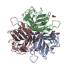
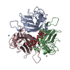
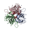
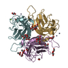
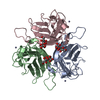
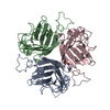
Mass: 21716.535 Da / Num. of mol.: 3 / Fragment: HEAD DOMAIN RESIDUES 172-365 / Mutation: YES
Source method: isolated from a genetically manipulated source
Details: ONLY THE KNOB DOMAIN, AFTER TEV CLEAVAGE OF A HIS-TAGGED PROTEIN CONSTRUCT
Source: (gene. exp.) HUMAN ADENOVIRUS TYPE 37 / Strain: P / Plasmid: PPROEX HTB / Production host: ESCHERICHIA COLI (E. coli) / Strain (production host): BL21(DE3) / References: UniProt: Q64823
#2: Chemical ChemComp-ZN / ZINC ION


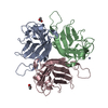
Mass: 65.409 Da / Num. of mol.: 3 / Source method: obtained synthetically / Formula: Zn
#3: Chemical ChemComp-ACT / ACETATE ION


Mass: 59.044 Da / Num. of mol.: 2 / Source method: obtained synthetically / Formula: C2H3O2
#4: Water ChemComp-HOH / water


Mass: 18.015 Da / Num. of mol.: 385 / Source method: isolated from a natural source / Formula: H2O
Compound detailsENGINEERED MUTATION IN CHAINS A-C, TYR 173 TO ALA 173 ENGINEERED MUTATION IN CHAINS A-C, LEU 174 TO ...ENGINEERED MUTATION IN CHAINS A-C, TYR 173 TO ALA 173 ENGINEERED MUTATION IN CHAINS A-C, LEU 174 TO MET 174 ENGINEERED MUTATION IN CHAINS A-C, VAL 175 TO GLY 175 ENGINEERED MUTATION IN CHAINS A-C, ALA 176 TO SER 176
Sequence detailsTHE FIRST FIVE RESIDUES HAVE BEEN CHANGED IN ORDER TO INTRODUCE THE TEV CLEAVAGE SITE

-
Experimental details

-
Experiment

ExperimentMethod: X-RAY DIFFRACTION / Number of used crystals: 1

-
Sample preparation

CrystalDensity Matthews: 2.43 Å3/Da / Density % sol: 49 % / Description: ISOMORPHOUS
Crystal growpH: 7.5
Details: RESERVOIR: 24 % PEG8000, 50 MM ZINC ACETATE, 100 MM HEPES, PH 7.5 PROTEIN: 30 MM TRIS-HCL, PH 7.5, 150 MM NACL

-
Data collection

DiffractionMean temperature: 100 K
Diffraction sourceSource: SYNCHROTRON / Site: ESRF / Beamline: ID29 / Wavelength: 0.979
DetectorType: ADSC CCD / Detector: CCD / Date: Apr 9, 2002 / Details: TOROIDAL MIRROR
RadiationMonochromator: SI(111) / Protocol: SINGLE WAVELENGTH / Monochromatic (M) / Laue (L): M / Scattering type: x-ray
Radiation wavelengthWavelength: 0.979 Å / Relative weight: 1
ReflectionResolution: 2→51.3 Å / Num. obs: 42295 / % possible obs: 98.2 % / Redundancy: 3.58 % / Biso Wilson estimate: 24.51 Å2 / Rmerge(I) obs: 0.107 / Net I/σ(I): 4.177
Reflection shellResolution: 2→2.11 Å / Redundancy: 3.32 % / Rmerge(I) obs: 0.25 / Mean I/σ(I) obs: 2.45 / % possible all: 96.9

-
Processing

Software
NameVersionClassification
CNS1refinement
MOSFLMdata reduction
SCALAdata scaling
RefinementMethod to determine structure: MOLECULAR REPLACEMENT
Starting model: PDB ENTRY 1UXA
Resolution: 2→51.3 Å / Rfactor Rfree error: 0.005 / Isotropic thermal model: RESTRAINED / Cross valid method: THROUGHOUT / σ(F): 0
Details: THE ELECTRON DENSITY THAT IS ASSOCIATED WITH THE ACETATE IONS IS POORLY DEFINED
RfactorNum. reflection% reflectionSelection details
Rfree0.241 2170 5 %RANDOM
Rwork0.204 ---
obs0.204 42597 96.5 %-
Solvent computationSolvent model: FLAT MODEL / Bsol: 35.04 Å2 / ksol: 0.322 e/Å3
Displacement parametersBiso mean: 27.13 Å2
Baniso -1Baniso -2Baniso -3
1--4.833 Å20 Å2-3.057 Å2
2---0.307 Å20 Å2
3---5.139 Å2
Refine analyze
FreeObs
Luzzati coordinate error0.3 Å0.25 Å
Luzzati d res low-5 Å
Luzzati sigma a0.29 Å0.26 Å
Refinement stepCycle: LAST / Resolution: 2→51.3 Å
ProteinNucleic acidLigandSolventTotal
Num. atoms4349 0 11 385 4745
Refine LS restraints
Refine-IDTypeDev idealDev ideal target
X-RAY DIFFRACTIONc_bond_d0.0228
X-RAY DIFFRACTIONc_bond_d_na
X-RAY DIFFRACTIONc_bond_d_prot
X-RAY DIFFRACTIONc_angle_d
X-RAY DIFFRACTIONc_angle_d_na
X-RAY DIFFRACTIONc_angle_d_prot
X-RAY DIFFRACTIONc_angle_deg2
X-RAY DIFFRACTIONc_angle_deg_na
X-RAY DIFFRACTIONc_angle_deg_prot
X-RAY DIFFRACTIONc_dihedral_angle_d26.97
X-RAY DIFFRACTIONc_dihedral_angle_d_na
X-RAY DIFFRACTIONc_dihedral_angle_d_prot
X-RAY DIFFRACTIONc_improper_angle_d1.23
X-RAY DIFFRACTIONc_improper_angle_d_na
X-RAY DIFFRACTIONc_improper_angle_d_prot
X-RAY DIFFRACTIONc_mcbond_it2.5593.5
X-RAY DIFFRACTIONc_mcangle_it3.3624
X-RAY DIFFRACTIONc_scbond_it3.3534
X-RAY DIFFRACTIONc_scangle_it4.4614.5
LS refinement shellResolution: 2→2.07 Å / Rfactor Rfree error: 0.03 / Total num. of bins used: 10
RfactorNum. reflection% reflection
Rfree0.309 210 5.6 %
Rwork0.286 3718 -
obs--89.74 %
Xplor file
Refine-IDSerial noParam fileTopol file
X-RAY DIFFRACTION1PROTEIN.PARAMPROTEIN.TOP
X-RAY DIFFRACTION2WATER.PARAMWATER.TOP
X-RAY DIFFRACTION3ION.TOP

+
About Yorodumi

-
News

-
Feb 9, 2022. New format data for meta-information of EMDB entries

New format data for meta-information of EMDB entries

  • Version 3 of the EMDB header file is now the official format.
  • The previous official version 1.9 will be removed from the archive.

Related info.:EMDB header

External links:wwPDB to switch to version 3 of the EMDB data model

-
Aug 12, 2020. Covid-19 info

Covid-19 info

URL: https://pdbjlc1.pdbj.org/emnavi/covid19.php

New page: Covid-19 featured information page in EM Navigator.

Related info.:Covid-19 info / Mar 5, 2020. Novel coronavirus structure data

+
Mar 5, 2020. Novel coronavirus structure data

Novel coronavirus structure data

Related info.:Yorodumi Speices / Aug 12, 2020. Covid-19 info

External links:COVID-19 featured content - PDBj / Molecule of the Month (242):Coronavirus Proteases

+
Jan 31, 2019. EMDB accession codes are about to change! (news from PDBe EMDB page)

EMDB accession codes are about to change! (news from PDBe EMDB page)

  • The allocation of 4 digits for EMDB accession codes will soon come to an end. Whilst these codes will remain in use, new EMDB accession codes will include an additional digit and will expand incrementally as the available range of codes is exhausted. The current 4-digit format prefixed with “EMD-” (i.e. EMD-XXXX) will advance to a 5-digit format (i.e. EMD-XXXXX), and so on. It is currently estimated that the 4-digit codes will be depleted around Spring 2019, at which point the 5-digit format will come into force.
  • The EM Navigator/Yorodumi systems omit the EMD- prefix.

Related info.:Q: What is EMD? / ID/Accession-code notation in Yorodumi/EM Navigator

External links:EMDB Accession Codes are Changing Soon! / Contact to PDBj

+
Jul 12, 2017. Major update of PDB

Major update of PDB

  • wwPDB released updated PDB data conforming to the new PDBx/mmCIF dictionary.
  • This is a major update changing the version number from 4 to 5, and with Remediation, in which all the entries are updated.
  • In this update, many items about electron microscopy experimental information are reorganized (e.g. em_software).
  • Now, EM Navigator and Yorodumi are based on the updated data.

External links:wwPDB Remediation / Enriched Model Files Conforming to OneDep Data Standards Now Available in the PDB FTP Archive

-
Yorodumi

Thousand views of thousand structures

  • Yorodumi is a browser for structure data from EMDB, PDB, SASBDB, etc.
  • This page is also the successor to EM Navigator detail page, and also detail information page/front-end page for Omokage search.
  • The word "yorodu" (or yorozu) is an old Japanese word meaning "ten thousand". "mi" (miru) is to see.

Related info.:EMDB / PDB / SASBDB / Comparison of 3 databanks / Yorodumi Search / Aug 31, 2016. New EM Navigator & Yorodumi / Yorodumi Papers / Jmol/JSmol / Function and homology information / Changes in new EM Navigator and Yorodumi

Read more