 Movie
Movie Controller

[English] 日本語
 Yorodumi
- PDB-1usr: Newcastle disease virus hemagglutinin-neuraminidase: Evidence for... -
+ Open data

- Basic information
| Entry | Database: PDB / ID: 1usr | ||||||||||||
|---|---|---|---|---|---|---|---|---|---|---|---|---|---|
| Title | Newcastle disease virus hemagglutinin-neuraminidase: Evidence for a second sialic acid binding site and implications for fusion | ||||||||||||
 Components Components | HEMAGGLUTININ-NEURAMINIDASE GLYCOPROTEIN | ||||||||||||
 Keywords Keywords | HYDROLASE / SIALIDASE / NEURAMINIDASE / HEMAGGLUTININ-NEURAMINIDASE / HEMAGGLUTININ / GLYCOPROTEIN | ||||||||||||
| Function / homology |  Function and homology information Function and homology informationexo-alpha-sialidase / exo-alpha-sialidase activity / host cell surface receptor binding / viral envelope / symbiont entry into host cell / virion attachment to host cell / host cell plasma membrane / virion membrane / identical protein binding Similarity search - Function | ||||||||||||
| Biological species |  NEWCASTLE DISEASE VIRUS NEWCASTLE DISEASE VIRUS | ||||||||||||
| Method |  X-RAY DIFFRACTION / SYNCHROTRON / MOLECULAR REPLACEMENT / Resolution: 2 Å X-RAY DIFFRACTION / SYNCHROTRON / MOLECULAR REPLACEMENT / Resolution: 2 Å | ||||||||||||
 Authors Authors | Zaitsev, V. / Von Itzstein, M. / Groves, D. / Kiefel, M. / Takimoto, T. / Portner, A. / Taylor, G. | ||||||||||||
 Citation Citation | Journal: J.Virol. / Year: 2004 Title: Second Sialic Acid Binding Site in Newcastle Disease Virus Hemagglutinin-Neuraminidase: Implications for Fusion Authors: Zaitsev, V. / Von Itzstein, M. / Groves, D. / Kiefel, M. / Takimoto, T. / Portner, A. / Taylor, G. #1: Journal: Nat.Struct.Biol. / Year: 2000Title: Crystal Structure of the Multifunctional Paramyxovirus Hemagglutinin-Neuraminidase Authors: Crennell, S. / Takimoto, T. / Portner, A. / Taylor, G. #2: Journal: Virology / Year: 2000 Title: Crystallization of Newcastle Disease Virus Hemagglutinin-Neuraminidase Glycoprotein Authors: Takimoto, T. / Taylor, G.L. / Crennell, S.J. / Scroggs, R.A. / Portner, A. | ||||||||||||
| History |
| ||||||||||||
| Remark 700 | SHEET THE SHEET STRUCTURE OF THIS MOLECULE IS BIFURCATED. IN ORDER TO REPRESENT THIS FEATURE IN ... SHEET THE SHEET STRUCTURE OF THIS MOLECULE IS BIFURCATED. IN ORDER TO REPRESENT THIS FEATURE IN THE SHEET RECORDS BELOW, TWO SHEETS ARE DEFINED. |
- Structure visualization
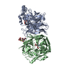
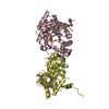
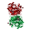
| Structure viewer | Molecule: MolmilJmol/JSmol |
|---|
- Downloads & links
-Download
| PDBx/mmCIF format | 1usr.cif.gz | 207.7 KB | Display | PDBx/mmCIF format |
|---|---|---|---|---|
| PDB format | pdb1usr.ent.gz | 162.4 KB | Display | PDB format |
| PDBx/mmJSON format | 1usr.json.gz | Tree view | PDBx/mmJSON format | |
| Others |  Other downloads Other downloads |
-Validation report
| Arichive directory | https://data.pdbj.org/pub/pdb/validation_reports/us/1usrftp://data.pdbj.org/pub/pdb/validation_reports/us/1usr | HTTPS FTP |
|---|
-Related structure data
| Related structure data |  1usxC 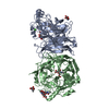 1e8vS C: citing same article ( S: Starting model for refinement |
|---|---|
| Similar structure data |


-Links
 PDBj
PDBj


- Assembly
| Deposited unit | 
| ||||||||
|---|---|---|---|---|---|---|---|---|---|
| 1 |
| ||||||||
| Unit cell |
| ||||||||
| Noncrystallographic symmetry (NCS) | NCS oper: (Code: given Matrix: (-0.40303, -0.20105, 0.89283), Vector: |
-Components
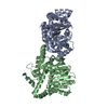
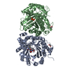
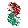
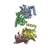
-Protein , 1 types, 2 molecules AB
| #1: Protein | Mass: 49856.910 Da / Num. of mol.: 2 / Fragment: HEAD DOMAIN, RESIDUES 124-577 / Source method: isolated from a natural source / Source: (natural) NEWCASTLE DISEASE VIRUS / Variant: KANSAS / References: UniProt: P32884, exo-alpha-sialidase |
|---|
-Sugars , 4 types, 6 molecules 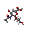


| #2: Polysaccharide | N-acetyl-alpha-neuraminic acid-(2-6)-methyl 6-thio-beta-D-galactopyranoside Type: oligosaccharide / Mass: 501.503 Da / Num. of mol.: 1 Source method: isolated from a genetically manipulated source | ||||
|---|---|---|---|---|---|
| #3: Sugar |  Type: D-saccharide / Mass: 291.255 Da / Num. of mol.: 2 / Source method: obtained synthetically / Formula: C11H17NO8 Type: D-saccharide / Mass: 291.255 Da / Num. of mol.: 2 / Source method: obtained synthetically / Formula: C11H17NO8#4: Sugar |  Type: D-saccharide, alpha linking / Mass: 221.208 Da / Num. of mol.: 2 Type: D-saccharide, alpha linking / Mass: 221.208 Da / Num. of mol.: 2Source method: isolated from a genetically manipulated source Formula: C8H15NO6 #6: Sugar | ChemComp-SIA / |  Type: D-saccharide, alpha linking / Mass: 309.270 Da / Num. of mol.: 1 Type: D-saccharide, alpha linking / Mass: 309.270 Da / Num. of mol.: 1Source method: isolated from a genetically manipulated source Formula: C11H19NO9 |
-Non-polymers , 2 types, 923 molecules 

| #5: Chemical |  Mass: 40.078 Da / Num. of mol.: 2 / Source method: obtained synthetically / Formula: Ca Mass: 40.078 Da / Num. of mol.: 2 / Source method: obtained synthetically / Formula: Ca#7: Water | ChemComp-HOH / | Mass: 18.015 Da / Num. of mol.: 921 / Source method: isolated from a natural source / Formula: H2O |
|---|
-Details
| Has protein modification | Y |
|---|
-Experimental details
-Experiment
| Experiment | Method: X-RAY DIFFRACTION / Number of used crystals: 1 |
|---|
- Sample preparation
| Crystal | Density Matthews: 2.9 Å3/Da / Density % sol: 57 % | |||||||||||||||||||||||||||||||||||||||||||||||||
|---|---|---|---|---|---|---|---|---|---|---|---|---|---|---|---|---|---|---|---|---|---|---|---|---|---|---|---|---|---|---|---|---|---|---|---|---|---|---|---|---|---|---|---|---|---|---|---|---|---|---|
| Crystal grow | pH: 6.5 / Details: pH 6.50 | |||||||||||||||||||||||||||||||||||||||||||||||||
| Crystal grow | *PLUS pH: 6.3 / Method: unknown | |||||||||||||||||||||||||||||||||||||||||||||||||
| Components of the solutions | *PLUS
|
-Data collection
| Diffraction | Mean temperature: 100 K |
|---|---|
| Diffraction source | Source: SYNCHROTRON / Site: SRS  / Beamline: PX14.2 / Wavelength: 0.979 / Beamline: PX14.2 / Wavelength: 0.979 |
| Detector | Type: ADSC CCD / Detector: CCD / Date: Oct 15, 2002 / Details: RH COATED SILICON MIRROR |
| Radiation | Monochromator: SILICON (111) MONOCHROMATORS / Protocol: SINGLE WAVELENGTH / Monochromatic (M) / Laue (L): M / Scattering type: x-ray |
| Radiation wavelength | Wavelength: 0.979 Å / Relative weight: 1 |
| Reflection | Resolution: 2→30 Å / Num. obs: 89733 / % possible obs: 98.5 % / Redundancy: 14.6 % / Biso Wilson estimate: 28.7 Å2 / Rmerge(I) obs: 0.083 / Net I/σ(I): 7.6 |
| Reflection shell | Resolution: 2→2.14 Å / Redundancy: 2.1 % / Rmerge(I) obs: 0.27 / Mean I/σ(I) obs: 3.2 / % possible all: 97 |
| Reflection | *PLUS Highest resolution: 2 Å / Num. measured all: 471352 / Rmerge(I) obs: 0.083 |
| Reflection shell | *PLUS % possible obs: 96.6 % / Rmerge(I) obs: 0.293 / Mean I/σ(I) obs: 2.8 |
- Processing
| Software |
| ||||||||||||||||||||||||||||||||||||||||||||||||||||||||||||
|---|---|---|---|---|---|---|---|---|---|---|---|---|---|---|---|---|---|---|---|---|---|---|---|---|---|---|---|---|---|---|---|---|---|---|---|---|---|---|---|---|---|---|---|---|---|---|---|---|---|---|---|---|---|---|---|---|---|---|---|---|---|
| Refinement | Method to determine structure: MOLECULAR REPLACEMENT Starting model: PDB ENTRY 1E8V Resolution: 2→12 Å / Data cutoff high absF: 1000 / Isotropic thermal model: RESTRAINED / Cross valid method: THROUGHOUT / σ(F): 0
| ||||||||||||||||||||||||||||||||||||||||||||||||||||||||||||
| Solvent computation | Solvent model: FLAT MODEL / Bsol: 60 Å2 / ksol: 0.41 e/Å3 | ||||||||||||||||||||||||||||||||||||||||||||||||||||||||||||
| Displacement parameters | Biso mean: 23.9 Å2
| ||||||||||||||||||||||||||||||||||||||||||||||||||||||||||||
| Refine analyze | Luzzati d res low obs: 12 Å | ||||||||||||||||||||||||||||||||||||||||||||||||||||||||||||
| Refinement step | Cycle: LAST / Resolution: 2→12 Å
| ||||||||||||||||||||||||||||||||||||||||||||||||||||||||||||
| Refine LS restraints |
| ||||||||||||||||||||||||||||||||||||||||||||||||||||||||||||
| LS refinement shell | Resolution: 2→2.1 Å / Total num. of bins used: 50 | ||||||||||||||||||||||||||||||||||||||||||||||||||||||||||||
| Refinement | *PLUS Rfactor Rfree: 0.227 / Rfactor Rwork: 0.191 | ||||||||||||||||||||||||||||||||||||||||||||||||||||||||||||
| Solvent computation | *PLUS | ||||||||||||||||||||||||||||||||||||||||||||||||||||||||||||
| Displacement parameters | *PLUS |
