 Movie
Movie Controller

[English] 日本語
 Yorodumi
- PDB-1urz: Low pH induced, membrane fusion conformation of the envelope prot... -
+ Open data

- Basic information
| Entry | Database: PDB / ID: 1urz | ||||||
|---|---|---|---|---|---|---|---|
| Title | Low pH induced, membrane fusion conformation of the envelope protein of tick-borne encephalitis virus | ||||||
 Components Components | ENVELOPE PROTEIN | ||||||
 Keywords Keywords | VIRUS/VIRAL PROTEIN / ENVELOPE PROTEIN / MEMBRANE FUSION / VIRUS-VIRAL PROTEIN complex | ||||||
| Function / homology |  Function and homology information Function and homology informationprotein dimerization activity / host cell endoplasmic reticulum membrane / fusion of virus membrane with host endosome membrane / symbiont entry into host cell / virion attachment to host cell / virion membrane / extracellular region / membrane Similarity search - Function | ||||||
| Biological species |  TICK-BORNE ENCEPHALITIS VIRUS TICK-BORNE ENCEPHALITIS VIRUS | ||||||
| Method |  X-RAY DIFFRACTION / SYNCHROTRON / MOLECULAR REPLACEMENT / Resolution: 2.7 Å X-RAY DIFFRACTION / SYNCHROTRON / MOLECULAR REPLACEMENT / Resolution: 2.7 Å | ||||||
 Authors Authors | Bressanelli, S. / Rey, F.A. | ||||||
 Citation Citation | Journal: Embo J. / Year: 2004 Title: Structure of a Flavivirus Envelope Glycoprotein in its Low-Ph-Induced Membrane Fusion Conformation. Authors: Bressanelli, S. / Stiasny, K. / Allison, S.L. / Stura, E.A. / Duquerroy, S. / Lescar, J. / Heinz, F.X. / Rey, F.A. | ||||||
| History |
| ||||||
| Remark 700 | SHEET THE SHEET STRUCTURE OF THIS MOLECULE IS BIFURCATED. IN ORDER TO REPRESENT THIS FEATURE IN ... SHEET THE SHEET STRUCTURE OF THIS MOLECULE IS BIFURCATED. IN ORDER TO REPRESENT THIS FEATURE IN THE SHEET RECORDS BELOW, TWO SHEETS ARE DEFINED. |
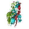
- Structure visualization
| Structure viewer | Molecule: MolmilJmol/JSmol |
|---|
- Downloads & links
-Download
| PDBx/mmCIF format | 1urz.cif.gz | 443.1 KB | Display | PDBx/mmCIF format |
|---|---|---|---|---|
| PDB format | pdb1urz.ent.gz | 365.4 KB | Display | PDB format |
| PDBx/mmJSON format | 1urz.json.gz | Tree view | PDBx/mmJSON format | |
| Others |  Other downloads Other downloads |
-Validation report
| Summary document | 1urz_validation.pdf.gz | 478.4 KB | Display | wwPDB validaton report |
|---|---|---|---|---|
| Full document | 1urz_full_validation.pdf.gz | 530.5 KB | Display | |
| Data in XML | 1urz_validation.xml.gz | 85.2 KB | Display | |
| Data in CIF | 1urz_validation.cif.gz | 116.2 KB | Display | |
| Arichive directory | https://data.pdbj.org/pub/pdb/validation_reports/ur/1urzftp://data.pdbj.org/pub/pdb/validation_reports/ur/1urz | HTTPS FTP |
-Related structure data
| Related structure data | 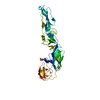 1svbS S: Starting model for refinement |
|---|---|
| Similar structure data |


-Links
 PDBj
PDBj

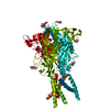
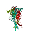
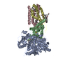
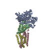
- Assembly
| Deposited unit | 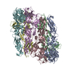
| ||||||||||||||||||||||||
|---|---|---|---|---|---|---|---|---|---|---|---|---|---|---|---|---|---|---|---|---|---|---|---|---|---|
| 1 | 
| ||||||||||||||||||||||||
| 2 | 
| ||||||||||||||||||||||||
| Unit cell |
| ||||||||||||||||||||||||
| Noncrystallographic symmetry (NCS) | NCS oper:
| ||||||||||||||||||||||||
| Details | THE VIRUS PARTICLE HAS A PROTEIN SHELL WITH ICOSAHEDRAL SYMMETRY MADE OF 180 DIMERS BEFORE EXPOSURE TO LOW PH. AFTER REARRANGEMENT AT LOW PH, THE SHELL LOSES ICOSAHEDRAL SYMMETRY AS THE ENVELOPE REARRANGES INTO 120 TRIMERS. |
-Components
| #1: Protein | Mass: 43789.758 Da / Num. of mol.: 6 / Source method: isolated from a natural source / Source: (natural) TICK-BORNE ENCEPHALITIS VIRUS / Strain: NEUDOERFL / References: UniProt: Q80E47#2: Water | ChemComp-HOH / |  Mass: 18.015 Da / Num. of mol.: 370 / Source method: isolated from a natural source / Formula: H2O Mass: 18.015 Da / Num. of mol.: 370 / Source method: isolated from a natural source / Formula: H2OHas protein modification | Y | |
|---|
-Experimental details
-Experiment
| Experiment | Method: X-RAY DIFFRACTION / Number of used crystals: 1 |
|---|
- Sample preparation
| Crystal | Density Matthews: 2.9 Å3/Da / Density % sol: 57 % Description: DATA CORRECTED FOR SPINDLE-SHUTTER DESYNCHRONISATION | ||||||||||||||||||||||||
|---|---|---|---|---|---|---|---|---|---|---|---|---|---|---|---|---|---|---|---|---|---|---|---|---|---|
| Crystal grow | pH: 4.5 Details: PEG 4000 25%, SODIUM ACETATE 0.1M PH 4.5, N,N-DIMETHYLDECYLAMINE N-OXIDE (DDAO) 15 MM | ||||||||||||||||||||||||
| Crystal grow | *PLUS Method: vapor diffusion, hanging drop / Details: Stiasny, K., (2004) J. Virol., 78, 3178. | ||||||||||||||||||||||||
| Components of the solutions | *PLUS
|
-Data collection
| Diffraction | Mean temperature: 100 K |
|---|---|
| Diffraction source | Source: SYNCHROTRON / Site: SLS  / Beamline: X06SA / Wavelength: 1.34106 / Beamline: X06SA / Wavelength: 1.34106 |
| Detector | Type: MARRESEARCH / Detector: CCD / Date: Jun 15, 2003 / Details: MIRRORS |
| Radiation | Monochromator: SI(111) / Protocol: SINGLE WAVELENGTH / Monochromatic (M) / Laue (L): M / Scattering type: x-ray |
| Radiation wavelength | Wavelength: 1.34106 Å / Relative weight: 1 |
| Reflection | Resolution: 2.7→40 Å / Num. obs: 80896 / % possible obs: 96.9 % / Observed criterion σ(I): -3 / Redundancy: 3.8 % / Biso Wilson estimate: 41.5 Å2 / Rmerge(I) obs: 0.111 / Net I/σ(I): 5.4 |
| Reflection shell | Resolution: 2.7→2.8 Å / Redundancy: 1.9 % / Rmerge(I) obs: 0.455 / Mean I/σ(I) obs: 2 / % possible all: 83.9 |
| Reflection | *PLUS Num. measured all: 306381 |
| Reflection shell | *PLUS % possible obs: 83.9 % |
- Processing
| Software |
| ||||||||||||||||||||||||||||||||||||||||||||||||||||||||||||||||||||||||||||||||
|---|---|---|---|---|---|---|---|---|---|---|---|---|---|---|---|---|---|---|---|---|---|---|---|---|---|---|---|---|---|---|---|---|---|---|---|---|---|---|---|---|---|---|---|---|---|---|---|---|---|---|---|---|---|---|---|---|---|---|---|---|---|---|---|---|---|---|---|---|---|---|---|---|---|---|---|---|---|---|---|---|---|
| Refinement | Method to determine structure: MOLECULAR REPLACEMENT Starting model: PDB ENTRY 1SVB Resolution: 2.7→40 Å / Rfactor Rfree error: 0.004 / Data cutoff high absF: 10000 / Isotropic thermal model: RESTRAINED / Cross valid method: THROUGHOUT / σ(F): 0 / Stereochemistry target values: MLF
| ||||||||||||||||||||||||||||||||||||||||||||||||||||||||||||||||||||||||||||||||
| Solvent computation | Solvent model: FLAT MODEL / Bsol: 24.1336 Å2 / ksol: 0.318351 e/Å3 | ||||||||||||||||||||||||||||||||||||||||||||||||||||||||||||||||||||||||||||||||
| Displacement parameters | Biso mean: 40.4 Å2
| ||||||||||||||||||||||||||||||||||||||||||||||||||||||||||||||||||||||||||||||||
| Refine analyze |
| ||||||||||||||||||||||||||||||||||||||||||||||||||||||||||||||||||||||||||||||||
| Refinement step | Cycle: LAST / Resolution: 2.7→40 Å
| ||||||||||||||||||||||||||||||||||||||||||||||||||||||||||||||||||||||||||||||||
| Refine LS restraints |
| ||||||||||||||||||||||||||||||||||||||||||||||||||||||||||||||||||||||||||||||||
| Refine LS restraints NCS | Rms dev Biso : 4.11 Å2 / Rms dev position: 0.36 Å / Weight Biso : 2 / Weight position: 10 | ||||||||||||||||||||||||||||||||||||||||||||||||||||||||||||||||||||||||||||||||
| LS refinement shell | Resolution: 2.7→2.87 Å / Rfactor Rfree error: 0.014 / Total num. of bins used: 6
| ||||||||||||||||||||||||||||||||||||||||||||||||||||||||||||||||||||||||||||||||
| Xplor file |
| ||||||||||||||||||||||||||||||||||||||||||||||||||||||||||||||||||||||||||||||||
| Refinement | *PLUS Rfactor Rwork: 0.206 | ||||||||||||||||||||||||||||||||||||||||||||||||||||||||||||||||||||||||||||||||
| Solvent computation | *PLUS | ||||||||||||||||||||||||||||||||||||||||||||||||||||||||||||||||||||||||||||||||
| Displacement parameters | *PLUS | ||||||||||||||||||||||||||||||||||||||||||||||||||||||||||||||||||||||||||||||||
| Refine LS restraints | *PLUS
| ||||||||||||||||||||||||||||||||||||||||||||||||||||||||||||||||||||||||||||||||
| LS refinement shell | *PLUS Highest resolution: 2.7 Å / Lowest resolution: 2.8 Å / Rfactor Rfree: 0.34 / Rfactor Rwork: 0.316 |
