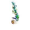
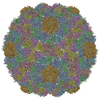
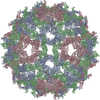
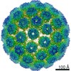
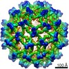
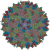
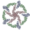
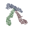
Journal: EMBO J / Year: 2003 Title: Structures of immature flavivirus particles. Authors: Ying Zhang / Jeroen Corver / Paul R Chipman / Wei Zhang / Sergei V Pletnev / Dagmar Sedlak / Timothy S Baker / James H Strauss / Richard J Kuhn / Michael G Rossmann / Abstract: Structures of prM-containing dengue and yellow fever virus particles were determined to 16 and 25 A resolution, respectively, by cryoelectron microscopy and image reconstruction techniques. The ...Structures of prM-containing dengue and yellow fever virus particles were determined to 16 and 25 A resolution, respectively, by cryoelectron microscopy and image reconstruction techniques. The closely similar structures show 60 icosahedrally organized trimeric spikes on the particle surface. Each spike consists of three prM:E heterodimers, where E is an envelope glycoprotein and prM is the precursor to the membrane protein M. The pre-peptide components of the prM proteins in each spike cover the fusion peptides at the distal ends of the E glycoproteins in a manner similar to the organization of the glycoproteins in the alphavirus spikes. Each heterodimer is associated with an E and a prM transmembrane density. These transmembrane densities represent either an EE or prMprM antiparallel coiled coil by which each protein spans the membrane twice, leaving the C-terminus of each protein on the exterior of the viral membrane, consistent with the predicted membrane-spanning domains of the unprocessed polyprotein.
SEQUENCE AUTHORS PROVIDED COORDINATES FOR ALPHA CARBONS ONLY. THE SEQRES RECORDS ARE FROM TICK-BORN ...SEQUENCE AUTHORS PROVIDED COORDINATES FOR ALPHA CARBONS ONLY. THE SEQRES RECORDS ARE FROM TICK-BORN ENCEPHALITIS VIRUS BECAUSE THE FITTING MODEL OF MAJOR ENVELOPE PROTEIN E (1SVB) WAS USED IN THIS STUDY.
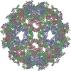
A: major envelope protein E B: major envelope protein E C: major envelope protein E
x 5
icosahedral pentamer
648 kDa, 15 polymers
Theoretical mass
Number of molelcules
Total (without water)
648,467
15
Polymers
648,467
15
Non-polymers
0
0
Water
0
Type
Name
Symmetry operation
Number
identity operation
1_555
x,y,z
1
point symmetry operation
4
4
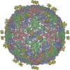
A: major envelope protein E B: major envelope protein E C: major envelope protein E
x 6
icosahedral 23 hexamer
778 kDa, 18 polymers
Theoretical mass
Number of molelcules
Total (without water)
778,161
18
Polymers
778,161
18
Non-polymers
0
0
Water
0
Type
Name
Symmetry operation
Number
identity operation
1_555
x,y,z
1
point symmetry operation
5
5
Idetical with deposited unit in distinct coordinate
icosahedral asymmetric unit, std point frame
Type
Name
Symmetry operation
Number
transform to point frame
1
Symmetry
Point symmetry: (Hermann–Mauguin notation: 532 / Schoenflies symbol: I (icosahedral))
-
Components
#1: Protein
majorenvelopeproteinE / Coordinate model: Cα atoms only
Mass: 43231.145 Da / Num. of mol.: 3 / Source method: isolated from a natural source / Source: (natural) Dengue virus 2 Puerto Rico/PR159-S1/1969 / Genus: Flavivirus / Species: Dengue virus / Strain: PR159/S1 / References: UniProt: P14336
-
Experimental details
-
Experiment
Experiment
Method: ELECTRON MICROSCOPY
EM experiment
Aggregation state: PARTICLE / 3D reconstruction method: single particle reconstruction
-
Sample preparation
Component
Name: dengue-2 immature particle / Type: COMPLEX Details: The samples were produced by adding ammonium cloride to the medium in the late infection stage
Specimen
Embedding applied: NO / Shadowing applied: NO / Staining applied: NO / Vitrification applied: YES
Crystal grow
*PLUS
Method: electron microscopy / Details: electron microscopy
-
Electron microscopy imaging
Microscopy
Model: FEI/PHILIPS CM300FEG/T
Electron gun
Electron source: FIELD EMISSION GUN / Illumination mode: FLOOD BEAM
Electron lens
Mode: BRIGHT FIELD / Nominal magnification: 33000 X / Nominal defocus max: 3640 nm / Nominal defocus min: 1662 nm / Cs: 2 mm
Electron dose: 15 e/Å2 / Film or detector model: KODAK SO-163 FILM
-
Processing
EM software
ID
Name
Category
Details (eV)
1
EMfit
modelfitting
2
EMfit
modelfitting
3
Custom
3Dreconstruction
common-line method
CTF correction
Details: EACH VIRAL IMAGE WAS CTF CORRECTED BEFORE RECONSTRUCTION, BASED ON THE FOLLOWING EQUATION: F(CORR)=F(OBS)/[|CTF|+WIENER]
Symmetry
Point symmetry: I (icosahedral)
3D reconstruction
Method: COMMON-LINES AND POLAR-FOURIER-TRANSFORM ((FULLER ET AL. 1996,J.STRUC.BIOL. 116, 48-55 BAKER ANDCHENG, 1996, J.STRUC.BIOL. 116, 120-130) Resolution: 16 Å / Nominal pixel size: 4.24 Å / Symmetry type: POINT
Atomic model building
Protocol: OTHER / Space: REAL Details: METHOD--The atomic structure of TBEV-E monomer was first fitted in one position in one asymmetry unit, then the densities at all pixels covered by the first fitted monomer was set to zero. ...Details: METHOD--The atomic structure of TBEV-E monomer was first fitted in one position in one asymmetry unit, then the densities at all pixels covered by the first fitted monomer was set to zero. After that, the second monomer was fitted into the left densities. The densities coresponding to the second monomer was again set to zero, then the third monomer was fitted.
In the structure databanks used in Yorodumi, some data are registered as the other names, "COVID-19 virus" and "2019-nCoV". Here are the details of the virus and the list of structure data.
Jan 31, 2019. EMDB accession codes are about to change! (news from PDBe EMDB page)
EMDB accession codes are about to change! (news from PDBe EMDB page)
The allocation of 4 digits for EMDB accession codes will soon come to an end. Whilst these codes will remain in use, new EMDB accession codes will include an additional digit and will expand incrementally as the available range of codes is exhausted. The current 4-digit format prefixed with “EMD-” (i.e. EMD-XXXX) will advance to a 5-digit format (i.e. EMD-XXXXX), and so on. It is currently estimated that the 4-digit codes will be depleted around Spring 2019, at which point the 5-digit format will come into force.
The EM Navigator/Yorodumi systems omit the EMD- prefix.
Related info.:Q: What is EMD? / ID/Accession-code notation in Yorodumi/EM Navigator
Yorodumi is a browser for structure data from EMDB, PDB, SASBDB, etc.
This page is also the successor to EM Navigator detail page, and also detail information page/front-end page for Omokage search.
The word "yorodu" (or yorozu) is an old Japanese word meaning "ten thousand". "mi" (miru) is to see.
Related info.:EMDB / PDB / SASBDB / Comparison of 3 databanks / Yorodumi Search / Aug 31, 2016. New EM Navigator & Yorodumi / Yorodumi Papers / Jmol/JSmol / Function and homology information / Changes in new EM Navigator and Yorodumi
 Movie
Movie Controller


 Open data
 Basic information
 Components
 Keywords
 Function and homology information
 Dengue virus 2 Puerto Rico/PR159-S1/1969
 Authors
 Citation
 Structure visualization
 Downloads & links
 Other downloads
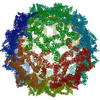
 PDBj
PDBj


 Assembly


 Sample preparation
 Electron microscopy imaging
 FIELD EMISSION GUN / Illumination mode: FLOOD BEAM
 Processing