 Movie
Movie Controller


+ Open data

- Basic information
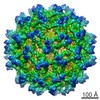
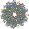
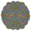
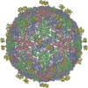
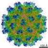
| Entry | Database: PDB / ID: 1tge | ||||||
|---|---|---|---|---|---|---|---|
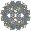
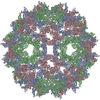
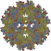
| Title | The structure of immature Dengue virus at 12.5 angstrom | ||||||
 Components Components | envelope glycoprotein | ||||||
 Keywords Keywords | VIRUS / flavivirus / dengue immature virus / prM particle / Icosahedral virus | ||||||
| Function / homology |  Function and homology information Function and homology informationflavivirin / serine-type peptidase activity / nucleoside-triphosphate phosphatase / double-stranded RNA binding / viral nucleocapsid / clathrin-dependent endocytosis of virus by host cell / RNA helicase activity / protein dimerization activity / host cell endoplasmic reticulum membrane / RNA helicase ...flavivirin / serine-type peptidase activity / nucleoside-triphosphate phosphatase / double-stranded RNA binding / viral nucleocapsid / clathrin-dependent endocytosis of virus by host cell / RNA helicase activity / protein dimerization activity / host cell endoplasmic reticulum membrane / RNA helicase / fusion of virus membrane with host endosome membrane / viral envelope / virion attachment to host cell / virion membrane / ATP hydrolysis activity / proteolysis / extracellular region / ATP binding Similarity search - Function | ||||||
| Biological species |  Dengue virus 2 Puerto Rico/PR159-S1/1969 Dengue virus 2 Puerto Rico/PR159-S1/1969 | ||||||
| Method | ELECTRON MICROSCOPY / single particle reconstruction / cryo EM / Resolution: 12.5 Å | ||||||
 Authors Authors | Zhang, Y. / Zhang, W. / Ogata, S. / Clements, D. / Strauss, J.H. / Baker, T.S. / Kuhn, R.J. / Rossmann, M.G. | ||||||
 Citation Citation | Journal: Structure / Year: 2004 Title: Conformational changes of the flavivirus E glycoprotein. Authors: Ying Zhang / Wei Zhang / Steven Ogata / David Clements / James H Strauss / Timothy S Baker / Richard J Kuhn / Michael G Rossmann /  Abstract: Dengue virus, a member of the Flaviviridae family, has a surface composed of 180 copies each of the envelope (E) glycoprotein and the membrane (M) protein. The crystal structure of an N-terminal ...Dengue virus, a member of the Flaviviridae family, has a surface composed of 180 copies each of the envelope (E) glycoprotein and the membrane (M) protein. The crystal structure of an N-terminal fragment of E has been determined and compared with a previously described structure. The primary difference between these structures is a 10 degrees rotation about a hinge relating the fusion domain DII to domains DI and DIII. These two rigid body components were used for independent fitting of E into the cryo-electron microscopy maps of both immature and mature dengue viruses. The fitted E structures in these two particles showed a difference of 27 degrees between the two components. Comparison of the E structure in its postfusion state with that in the immature and mature virions shows a rotation approximately around the same hinge. Flexibility of E is apparently a functional requirement for assembly and infection of flaviviruses. | ||||||
| History |
| ||||||
| Remark 999 | SEQUENCE AUTHORS SUBMITTED COORDINATES FOR CA ATOMS ONLY. |
- Structure visualization
| Movie |
Movie viewer |
|---|---|
| Structure viewer | Molecule: MolmilJmol/JSmol |
- Downloads & links
-Download
| PDBx/mmCIF format | 1tge.cif.gz | 48.5 KB | Display | PDBx/mmCIF format |
|---|---|---|---|---|
| PDB format | pdb1tge.ent.gz | 29.5 KB | Display | PDB format |
| PDBx/mmJSON format | 1tge.json.gz | Tree view | PDBx/mmJSON format | |
| Others |  Other downloads Other downloads |
-Validation report
| Arichive directory | https://data.pdbj.org/pub/pdb/validation_reports/tg/1tgeftp://data.pdbj.org/pub/pdb/validation_reports/tg/1tge | HTTPS FTP |
|---|
-Related structure data
| Related structure data |  5422MC 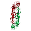 1tg8C  1thdC C: citing same article ( M: map data used to model this data |
|---|---|
| Similar structure data |


-Links
 PDBj
PDBj

- Assembly
| Deposited unit | 
|
|---|---|
| 1 | x 60
|
| 2 |
|
| 3 | x 5
|
| 4 | x 6
|
| 5 | 
|
| Symmetry | Point symmetry: (Hermann–Mauguin notation: 532 / Schoenflies symbol: I (icosahedral)) |
-Components
| #1: Protein | Mass: 43876.441 Da / Num. of mol.: 3 / Source method: isolated from a natural source / Source: (natural) Dengue virus 2 Puerto Rico/PR159-S1/1969 / Genus: Flavivirus / Species: Dengue virus / Strain: PR159/S1 / References: UniProt: P27914 |
|---|
-Experimental details
-Experiment
| Experiment | Method: ELECTRON MICROSCOPY |
|---|---|
| EM experiment | Aggregation state: PARTICLE / 3D reconstruction method: single particle reconstruction |
- Sample preparation
| Component | Name: Dengue-2 immature particle / Type: VIRUS Details: The samples were produced by adding ammonium chloride to the media in the late infection stage |
|---|---|
| Specimen | Embedding applied: NO / Shadowing applied: NO / Staining applied: NO / Vitrification applied: YES |
- Electron microscopy imaging
| Microscopy | Model: FEI/PHILIPS CM300FEG/T |
|---|---|
| Electron gun | Electron source:  FIELD EMISSION GUN / Illumination mode: FLOOD BEAM FIELD EMISSION GUN / Illumination mode: FLOOD BEAM |
| Electron lens | Mode: BRIGHT FIELD / Calibrated magnification: 33000 X / Nominal defocus max: 3640 nm / Nominal defocus min: 1662 nm / Cs: 2 mm |
| Specimen holder | Temperature: 100 K / Tilt angle max: 0 ° / Tilt angle min: 0 ° |
| Image recording | Electron dose: 15 e/Å2 / Film or detector model: KODAK SO-163 FILM |
- Processing
| EM software |
| ||||||||||||
|---|---|---|---|---|---|---|---|---|---|---|---|---|---|
| Symmetry | Point symmetry: I (icosahedral) | ||||||||||||
| 3D reconstruction | Method: See primary citation / Resolution: 12.5 Å / Symmetry type: POINT | ||||||||||||
| Atomic model building | Protocol: OTHER / Space: REAL / Details: METHOD--see primary citation | ||||||||||||
| Atomic model building | PDB-ID: 1TG8 Accession code: 1TG8 / Source name: PDB / Type: experimental model | ||||||||||||
| Refinement step | Cycle: LAST
|
