 Movie
Movie Controller

[English] 日本語
 Yorodumi
- PDB-1urx: Crystallographic structure of beta-agarase A in complex with olig... -
+ Open data

- Basic information
| Entry | Database: PDB / ID: 1urx | |||||||||
|---|---|---|---|---|---|---|---|---|---|---|
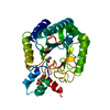
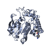
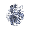
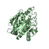
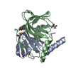
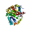
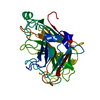
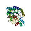
| Title | Crystallographic structure of beta-agarase A in complex with oligoagarose | |||||||||
 Components Components | BETA-AGARASE A | |||||||||
 Keywords Keywords | HYDROLASE / BETA-AGARASE / AGAROSE / GLYCOSIDE HYDROLASE / FAMILY 16 / DOUBLE HELIX / TWO BINDING-SITES | |||||||||
| Function / homology |  Function and homology information Function and homology informationbeta-agarase / beta-agarase activity / carbohydrate metabolic process / extracellular region Similarity search - Function | |||||||||
| Biological species |  ZOBELLIA GALACTANIVORANS (bacteria) ZOBELLIA GALACTANIVORANS (bacteria) | |||||||||
| Method |  X-RAY DIFFRACTION / SYNCHROTRON / MOLECULAR REPLACEMENT / Resolution: 1.7 Å X-RAY DIFFRACTION / SYNCHROTRON / MOLECULAR REPLACEMENT / Resolution: 1.7 Å | |||||||||
 Authors Authors | Allouch, J. / Helbert, W. / Henrissat, B. / Czjzek, M. | |||||||||
 Citation Citation | Journal: Structure / Year: 2004 Title: Parallel Substrate Binding Sites in a Beta-Agarase Suggest a Novel Mode of Action on Double-Helical Agarose Authors: Allouch, J. / Helbert, W. / Henrissat, B. / Czjzek, M. | |||||||||
| History |
| |||||||||
| Remark 700 | SHEET THE SHEET STRUCTURE OF THIS MOLECULE IS BIFURCATED. IN ORDER TO REPRESENT THIS FEATURE IN ... SHEET THE SHEET STRUCTURE OF THIS MOLECULE IS BIFURCATED. IN ORDER TO REPRESENT THIS FEATURE IN THE SHEET RECORDS BELOW, TWO SHEETS ARE DEFINED. |
- Structure visualization
| Structure viewer | Molecule: MolmilJmol/JSmol |
|---|
- Downloads & links
-Download
| PDBx/mmCIF format | 1urx.cif.gz | 82.1 KB | Display | PDBx/mmCIF format |
|---|---|---|---|---|
| PDB format | pdb1urx.ent.gz | 58.1 KB | Display | PDB format |
| PDBx/mmJSON format | 1urx.json.gz | Tree view | PDBx/mmJSON format | |
| Others |  Other downloads Other downloads |
-Validation report
| Arichive directory | https://data.pdbj.org/pub/pdb/validation_reports/ur/1urxftp://data.pdbj.org/pub/pdb/validation_reports/ur/1urx | HTTPS FTP |
|---|
-Related structure data
| Related structure data | 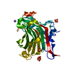 1o4yS S: Starting model for refinement |
|---|---|
| Similar structure data |


-Links
 PDBj
PDBj
- Assembly
| Deposited unit | 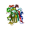
| ||||||||
|---|---|---|---|---|---|---|---|---|---|
| 1 |
| ||||||||
| Unit cell |
|
-Components
| #1: Protein | Mass: 32135.389 Da / Num. of mol.: 1 / Fragment: BETA-AGARASE A DOMAIN, RESIDUES 20-290 / Mutation: YES Source method: isolated from a genetically manipulated source Details: TWO MOLECULES OF OLIGOAGAROSE / Source: (gene. exp.) ZOBELLIA GALACTANIVORANS (bacteria) / Production host: References: UniProt: Q9RGX9, UniProt: G0L322*PLUS, beta-agarase |
|---|---|
| #2: Polysaccharide | 3,6-anhydro-alpha-L-galactopyranose-(1-3)-beta-D-galactopyranose-(1-4)-3,6-anhydro-alpha-L- ...3,6-anhydro-alpha-L-galactopyranose-(1-3)-beta-D-galactopyranose-(1-4)-3,6-anhydro-alpha-L-galactopyranose-(1-3)-alpha-D-galactopyranose Source method: isolated from a genetically manipulated source |
| #3: Polysaccharide | 3,6-anhydro-alpha-L-galactopyranose-(1-3)-beta-D-galactopyranose-(1-4)-3,6-anhydro-alpha-L- ...3,6-anhydro-alpha-L-galactopyranose-(1-3)-beta-D-galactopyranose-(1-4)-3,6-anhydro-alpha-L-galactopyranose-(1-3)-beta-D-galactopyranose-(1-4)-3,6-anhydro-alpha-L-galactopyranose-(1-3)-beta-D-galactopyranose-(1-4)-3,6-anhydro-alpha-L-galactopyranose Source method: isolated from a genetically manipulated source |
| #4: Chemical | ChemComp-CA /   Mass: 40.078 Da / Num. of mol.: 1 / Source method: obtained synthetically / Formula: Ca Mass: 40.078 Da / Num. of mol.: 1 / Source method: obtained synthetically / Formula: Ca |
| #5: Water | ChemComp-HOH /  Mass: 18.015 Da / Num. of mol.: 354 / Source method: isolated from a natural source / Formula: H2O Mass: 18.015 Da / Num. of mol.: 354 / Source method: isolated from a natural source / Formula: H2O |
| Compound details | ENGINEERED |
-Experimental details
-Experiment
| Experiment | Method: X-RAY DIFFRACTION / Number of used crystals: 1 |
|---|
- Sample preparation
| Crystal | Density Matthews: 2.81 Å3/Da / Density % sol: 56 % | ||||||||||||||||||||||||||||||||||||||||||||||||||||||||
|---|---|---|---|---|---|---|---|---|---|---|---|---|---|---|---|---|---|---|---|---|---|---|---|---|---|---|---|---|---|---|---|---|---|---|---|---|---|---|---|---|---|---|---|---|---|---|---|---|---|---|---|---|---|---|---|---|---|
| Crystal grow | Temperature: 293.65 K / Method: vapor diffusion, hanging drop / pH: 4.6 Details: 30 % PEG 4000 200 MM AMMONIUM ACETATE, 100 MM SODIUM ACETATE PH 4.6 | ||||||||||||||||||||||||||||||||||||||||||||||||||||||||
| Crystal grow | *PLUS Temperature: 20.5 ℃ / pH: 7.5 / Method: vapor diffusion, hanging drop | ||||||||||||||||||||||||||||||||||||||||||||||||||||||||
| Components of the solutions | *PLUS
|
-Data collection
| Diffraction | Mean temperature: 193 K |
|---|---|
| Diffraction source | Source: SYNCHROTRON / Site: ESRF  / Beamline: ID14-1 / Wavelength: 0.934 / Beamline: ID14-1 / Wavelength: 0.934 |
| Detector | Date: Mar 15, 2003 |
| Radiation | Protocol: SINGLE WAVELENGTH / Monochromatic (M) / Laue (L): M / Scattering type: x-ray |
| Radiation wavelength | Wavelength: 0.934 Å / Relative weight: 1 |
| Reflection | Resolution: 1.7→37.2 Å / Num. obs: 34045 / % possible obs: 95.3 % / Redundancy: 4.4 % / Rmerge(I) obs: 0.042 / Net I/σ(I): 12.1 |
| Reflection shell | Resolution: 1.7→1.76 Å / Redundancy: 4.5 % / Rmerge(I) obs: 0.11 / Mean I/σ(I) obs: 6.8 / % possible all: 98.4 |
| Reflection | *PLUS Highest resolution: 1.7 Å / Lowest resolution: 37.2 Å / Redundancy: 4.4 % / Num. measured all: 148960 / Rmerge(I) obs: 0.042 |
| Reflection shell | *PLUS % possible obs: 98.4 % / Redundancy: 4.5 % / Rmerge(I) obs: 0.11 / Mean I/σ(I) obs: 6.8 |
- Processing
| Software |
| ||||||||||||||||||||
|---|---|---|---|---|---|---|---|---|---|---|---|---|---|---|---|---|---|---|---|---|---|
| Refinement | Method to determine structure: MOLECULAR REPLACEMENT Starting model: PDB ENTRY 1O4Y Resolution: 1.7→37.2 Å / SU B: 1.641 / SU ML: 0.056 / Cross valid method: THROUGHOUT / ESU R: 0.095 / ESU R Free: 0.092
| ||||||||||||||||||||
| Displacement parameters | Biso mean: 13.555 Å2
| ||||||||||||||||||||
| Refinement step | Cycle: LAST / Resolution: 1.7→37.2 Å
| ||||||||||||||||||||
| Refinement | *PLUS Num. reflection obs: 32767 / % reflection Rfree: 5 % / Rfactor Rfree: 0.182 / Rfactor Rwork: 0.155 | ||||||||||||||||||||
| Solvent computation | *PLUS | ||||||||||||||||||||
| Displacement parameters | *PLUS | ||||||||||||||||||||
| Refine LS restraints | *PLUS
|
