 Movie
Movie Controller


+ Open data

- Basic information
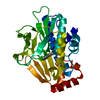
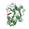
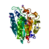
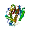
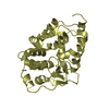
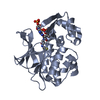
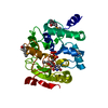
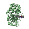
| Entry | Database: PDB / ID: 4bvl | ||||||
|---|---|---|---|---|---|---|---|
| Title | Structure of 202-208 deletion mutant of PhaZ7 PHB depolymerase | ||||||
 Components Components | PHB DEPOLYMERASE PHAZ7 | ||||||
 Keywords Keywords | HYDROLASE / CATALYTIC TRIAD / BIOPOLYMER BINDING | ||||||
| Function / homology |  Function and homology information Function and homology information | ||||||
| Biological species |  PAUCIMONAS LEMOIGNEI (bacteria) PAUCIMONAS LEMOIGNEI (bacteria) | ||||||
| Method |  X-RAY DIFFRACTION / SYNCHROTRON / MOLECULAR REPLACEMENT / Resolution: 2.002 Å X-RAY DIFFRACTION / SYNCHROTRON / MOLECULAR REPLACEMENT / Resolution: 2.002 Å | ||||||
 Authors Authors | Hermawan, S. / Subedi, B. / Papageorgiou, A.C. / Jendrossek, D. | ||||||
 Citation Citation | Journal: Mol.Microbiol. / Year: 2013 Title: Biochemical Analysis and Structure Determination of Paucimonas Lemoignei Poly(3-Hydroxybutyrate) (Phb) Depolymerase Phaz7 Muteins Reveal the Phb Binding Site and Details of Substrate-Enzyme Interactions. Authors: Jendrossek, D. / Hermawan, S. / Subedi, B. / Papageorgiou, A.C. #1: Journal: J.Mol.Biol. / Year: 2008Title: Structural Basis of Poly(3-Hydroxybutyrate) Hydrolysis by Phaz7 Depolymerase from Paucimonas Lemoignei. Authors: Papageorgiou, A.C. / Hermawan, S. / Singh, C.B. / Jendrossek, D. | ||||||
| History |
|
- Structure visualization
| Structure viewer | Molecule: MolmilJmol/JSmol |
|---|
- Downloads & links
-Download
| PDBx/mmCIF format | 4bvl.cif.gz | 290.4 KB | Display | PDBx/mmCIF format |
|---|---|---|---|---|
| PDB format | pdb4bvl.ent.gz | 236.1 KB | Display | PDB format |
| PDBx/mmJSON format | 4bvl.json.gz | Tree view | PDBx/mmJSON format | |
| Others |  Other downloads Other downloads |
-Validation report
| Arichive directory | https://data.pdbj.org/pub/pdb/validation_reports/bv/4bvlftp://data.pdbj.org/pub/pdb/validation_reports/bv/4bvl | HTTPS FTP |
|---|
-Related structure data
| Related structure data | 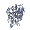 4brsSC 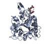 4btvC 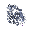 4bvjC 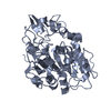 4bvkC 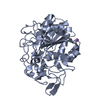 4bymC C: citing same article ( S: Starting model for refinement |
|---|---|
| Similar structure data |


-Links
 PDBj
PDBj- Assembly
| Deposited unit | 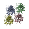
| ||||||||||||
|---|---|---|---|---|---|---|---|---|---|---|---|---|---|
| 1 | 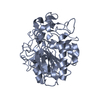
| ||||||||||||
| 2 | 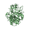
| ||||||||||||
| 3 | 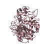
| ||||||||||||
| 4 | 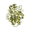
| ||||||||||||
| Unit cell |
| ||||||||||||
| Components on special symmetry positions |
|
-Components
| #1: Protein | Mass: 35282.934 Da / Num. of mol.: 4 / Fragment: RESIDUES 39-380 Source method: isolated from a genetically manipulated source Source: (gene. exp.) PAUCIMONAS LEMOIGNEI (bacteria) / Production host: References: UniProt: Q939Q9, poly(3-hydroxybutyrate) depolymerase #2: Water | ChemComp-HOH / |  Mass: 18.015 Da / Num. of mol.: 1803 / Source method: isolated from a natural source / Formula: H2O Mass: 18.015 Da / Num. of mol.: 1803 / Source method: isolated from a natural source / Formula: H2OHas protein modification | Y | Sequence details | RESIDUES 202-208 HAVE BEEN DELETED | |
|---|
-Experimental details
-Experiment
| Experiment | Method: X-RAY DIFFRACTION / Number of used crystals: 1 |
|---|
- Sample preparation
| Crystal | Density Matthews: 2 Å3/Da / Density % sol: 38 % / Description: NONE |
|---|---|
| Crystal grow | Method: vapor diffusion, hanging drop / pH: 5 Details: HANGING DROP VAPOUR DIFFUSION. PROTEIN CONCENTRATION 10 MG/ML. 0.1 M NAOAC (PH 5.0), 0.2 M LICL, 15% W/V PEG6000 |
-Data collection
| Diffraction | Mean temperature: 100 K |
|---|---|
| Diffraction source | Source: SYNCHROTRON / Site: EMBL/DESY, HAMBURG  / Beamline: X13 / Wavelength: 0.8123 / Beamline: X13 / Wavelength: 0.8123 |
| Detector | Type: MARRESEARCH SX-165 / Detector: CCD / Date: Nov 19, 2010 |
| Radiation | Protocol: SINGLE WAVELENGTH / Monochromatic (M) / Laue (L): M / Scattering type: x-ray |
| Radiation wavelength | Wavelength: 0.8123 Å / Relative weight: 1 |
| Reflection | Resolution: 2→19.6 Å / Num. obs: 74317 / % possible obs: 98.7 % / Observed criterion σ(I): -3 / Redundancy: 5.2 % / Biso Wilson estimate: 12.3 Å2 / Rmerge(I) obs: 0.11 / Net I/σ(I): 12.5 |
| Reflection shell | Resolution: 2→2.12 Å / Redundancy: 5.2 % / Rmerge(I) obs: 0.33 / Mean I/σ(I) obs: 4.4 / % possible all: 94.9 |
- Processing
| Software |
| ||||||||||||||||||||||||||||||||||||||||||||||||||||||||||||||||||||||||||||||||||||||||||||||||||||||||||||||||||||||||||||||||||||||||||||||||||||||||||||||||||||||||||||||||||||||||||||||||||||
|---|---|---|---|---|---|---|---|---|---|---|---|---|---|---|---|---|---|---|---|---|---|---|---|---|---|---|---|---|---|---|---|---|---|---|---|---|---|---|---|---|---|---|---|---|---|---|---|---|---|---|---|---|---|---|---|---|---|---|---|---|---|---|---|---|---|---|---|---|---|---|---|---|---|---|---|---|---|---|---|---|---|---|---|---|---|---|---|---|---|---|---|---|---|---|---|---|---|---|---|---|---|---|---|---|---|---|---|---|---|---|---|---|---|---|---|---|---|---|---|---|---|---|---|---|---|---|---|---|---|---|---|---|---|---|---|---|---|---|---|---|---|---|---|---|---|---|---|---|---|---|---|---|---|---|---|---|---|---|---|---|---|---|---|---|---|---|---|---|---|---|---|---|---|---|---|---|---|---|---|---|---|---|---|---|---|---|---|---|---|---|---|---|---|---|---|---|---|
| Refinement | Method to determine structure: MOLECULAR REPLACEMENT Starting model: PDB ENTRY 4BRS Resolution: 2.002→19.558 Å / SU ML: 0.17 / σ(F): 1.99 / Phase error: 17.37 / Stereochemistry target values: ML
| ||||||||||||||||||||||||||||||||||||||||||||||||||||||||||||||||||||||||||||||||||||||||||||||||||||||||||||||||||||||||||||||||||||||||||||||||||||||||||||||||||||||||||||||||||||||||||||||||||||
| Solvent computation | Shrinkage radii: 0.9 Å / VDW probe radii: 1.11 Å / Solvent model: FLAT BULK SOLVENT MODEL | ||||||||||||||||||||||||||||||||||||||||||||||||||||||||||||||||||||||||||||||||||||||||||||||||||||||||||||||||||||||||||||||||||||||||||||||||||||||||||||||||||||||||||||||||||||||||||||||||||||
| Displacement parameters | Biso mean: 12.7 Å2 | ||||||||||||||||||||||||||||||||||||||||||||||||||||||||||||||||||||||||||||||||||||||||||||||||||||||||||||||||||||||||||||||||||||||||||||||||||||||||||||||||||||||||||||||||||||||||||||||||||||
| Refinement step | Cycle: LAST / Resolution: 2.002→19.558 Å
| ||||||||||||||||||||||||||||||||||||||||||||||||||||||||||||||||||||||||||||||||||||||||||||||||||||||||||||||||||||||||||||||||||||||||||||||||||||||||||||||||||||||||||||||||||||||||||||||||||||
| Refine LS restraints |
| ||||||||||||||||||||||||||||||||||||||||||||||||||||||||||||||||||||||||||||||||||||||||||||||||||||||||||||||||||||||||||||||||||||||||||||||||||||||||||||||||||||||||||||||||||||||||||||||||||||
| LS refinement shell |
|
