 Movie
Movie Controller


+ Open data

- Basic information
| Entry | Database: PDB / ID: 3wdy | |||||||||
|---|---|---|---|---|---|---|---|---|---|---|
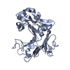
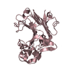
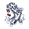
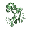
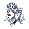
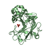
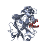
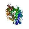
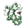
| Title | The complex structure of E113A with cellotetraose | |||||||||
 Components Components | Beta-1,3-1,4-glucanase | |||||||||
 Keywords Keywords | HYDROLASE / 1 / 3-1 / 4-beta-glucanase / 3(4)-beta-glucanase / PtLic16A / beta-jellyroll fold | |||||||||
| Function / homology |  Function and homology information Function and homology informationendo-1,3(4)-beta-glucanase / endo-1,3(4)-beta-glucanase activity / glucan catabolic process / side of membrane / plasma membrane Similarity search - Function | |||||||||
| Biological species |  Paecilomyces (fungus) Paecilomyces (fungus) | |||||||||
| Method |  X-RAY DIFFRACTION / SYNCHROTRON / MOLECULAR REPLACEMENT / Resolution: 1.94 Å X-RAY DIFFRACTION / SYNCHROTRON / MOLECULAR REPLACEMENT / Resolution: 1.94 Å | |||||||||
 Authors Authors | Cheng, Y.S. / Huang, C.H. / Chen, C.C. / Huang, T.Y. / Ko, T.P. / Huang, J.W. / Wu, T.H. / Liu, J.R. / Guo, R.T. | |||||||||
 Citation Citation | Journal: Biochim.Biophys.Acta / Year: 2014 Title: Structural and mutagenetic analyses of a 1,3-1,4-beta-glucanase from Paecilomyces thermophila Authors: Cheng, Y.S. / Huang, C.H. / Chen, C.C. / Huang, T.Y. / Ko, T.P. / Huang, J.W. / Wu, T.H. / Liu, J.R. / Guo, R.T. | |||||||||
| History |
|
- Structure visualization
| Structure viewer | Molecule: MolmilJmol/JSmol |
|---|
- Downloads & links
-Download
| PDBx/mmCIF format | 3wdy.cif.gz | 253 KB | Display | PDBx/mmCIF format |
|---|---|---|---|---|
| PDB format | pdb3wdy.ent.gz | 203.9 KB | Display | PDB format |
| PDBx/mmJSON format | 3wdy.json.gz | Tree view | PDBx/mmJSON format | |
| Others |  Other downloads Other downloads |
-Validation report
| Arichive directory | https://data.pdbj.org/pub/pdb/validation_reports/wd/3wdyftp://data.pdbj.org/pub/pdb/validation_reports/wd/3wdy | HTTPS FTP |
|---|
-Related structure data
| Related structure data |  3wdtSC  3wduC  3wdvC 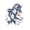 3wdwC  3wdxC S: Starting model for refinement C: citing same article ( |
|---|---|
| Similar structure data |

-Links
 PDBj
PDBj


- Assembly
| Deposited unit | 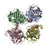
| ||||||||
|---|---|---|---|---|---|---|---|---|---|
| 1 | 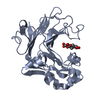
| ||||||||
| 2 | 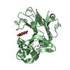
| ||||||||
| 3 | 
| ||||||||
| 4 | 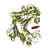
| ||||||||
| Unit cell |
|
-Components
-Protein , 1 types, 4 molecules ABCD
| #1: Protein | Mass: 31889.859 Da / Num. of mol.: 4 / Fragment: UNP residues 19-314 / Mutation: E113A Source method: isolated from a genetically manipulated source Source: (gene. exp.) Paecilomyces (fungus) / Strain: J18 / Plasmid: pPICZalphaA / Production host: Komagataella pastoris (fungus) / Strain (production host): X33 / References: UniProt: E0XN39, licheninase |
|---|
-Sugars , 3 types, 5 molecules 
| #2: Polysaccharide |   Source method: isolated from a genetically manipulated source Details: oligosaccharide / References: beta-cellobiose #3: Polysaccharide | beta-D-glucopyranose-(1-4)-beta-D-glucopyranose-(1-4)-beta-D-glucopyranose-(1-4)-beta-D-glucopyranose / beta-cellotetraose | 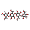  Source method: isolated from a genetically manipulated source Details: oligosaccharide / References: beta-cellotetraose #4: Sugar | ChemComp-BGC / |  Type: D-saccharide, beta linking / Mass: 180.156 Da / Num. of mol.: 1 Type: D-saccharide, beta linking / Mass: 180.156 Da / Num. of mol.: 1Source method: isolated from a genetically manipulated source Formula: C6H12O6 |
|---|
-Non-polymers , 2 types, 1005 molecules 

| #5: Chemical | ChemComp-SO4 /  Mass: 96.063 Da / Num. of mol.: 1 / Source method: obtained synthetically / Formula: SO4 Mass: 96.063 Da / Num. of mol.: 1 / Source method: obtained synthetically / Formula: SO4 |
|---|---|
| #6: Water | ChemComp-HOH / Mass: 18.015 Da / Num. of mol.: 1004 / Source method: isolated from a natural source / Formula: H2O |
-Details
| Has protein modification | Y |
|---|
-Experimental details
-Experiment
| Experiment | Method: X-RAY DIFFRACTION / Number of used crystals: 1 |
|---|
- Sample preparation
| Crystal | Density Matthews: 2.63 Å3/Da / Density % sol: 53.28 % |
|---|---|
| Crystal grow | Temperature: 298 K / Method: vapor diffusion, sitting drop / pH: 6.5 Details: 0.2M ammonium sulfate, 0.1M sodium cacodylate, 34%(w/v) PEG8000, pH 6.5, VAPOR DIFFUSION, SITTING DROP, temperature 298K |
-Data collection
| Diffraction | Mean temperature: 100 K |
|---|---|
| Diffraction source | Source: SYNCHROTRON / Site: NSRRC  / Beamline: BL13B1 / Wavelength: 1 Å / Beamline: BL13B1 / Wavelength: 1 Å |
| Detector | Type: ADSC QUANTUM 315r / Detector: CCD / Date: Oct 30, 2012 |
| Radiation | Protocol: SINGLE WAVELENGTH / Monochromatic (M) / Laue (L): M / Scattering type: x-ray |
| Radiation wavelength | Wavelength: 1 Å / Relative weight: 1 |
| Reflection | Resolution: 1.94→25 Å / Num. obs: 97796 / % possible obs: 99.9 % / Redundancy: 4.9 % / Rmerge(I) obs: 0.065 / Net I/σ(I): 20.3 |
| Reflection shell | Resolution: 1.94→2.01 Å / Redundancy: 4.9 % / Rmerge(I) obs: 0.443 / Mean I/σ(I) obs: 2.4 / Num. unique all: 9735 / % possible all: 99.9 |
- Processing
| Software |
| |||||||||||||||||||||||||
|---|---|---|---|---|---|---|---|---|---|---|---|---|---|---|---|---|---|---|---|---|---|---|---|---|---|---|
| Refinement | Method to determine structure: MOLECULAR REPLACEMENT Starting model: 3WDT Resolution: 1.94→24.67 Å / σ(F): 2 / Stereochemistry target values: Engh & Huber
| |||||||||||||||||||||||||
| Solvent computation | Bsol: 41.8041 Å2 | |||||||||||||||||||||||||
| Displacement parameters | Biso mean: 29.2327 Å2
| |||||||||||||||||||||||||
| Refinement step | Cycle: LAST / Resolution: 1.94→24.67 Å
| |||||||||||||||||||||||||
| Refine LS restraints |
| |||||||||||||||||||||||||
| LS refinement shell | Resolution: 1.94→2.01 Å /
|
