 Movie
Movie Controller


+ Open data

- Basic information
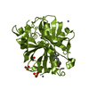
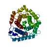
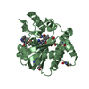
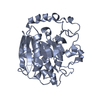
| Entry | Database: PDB / ID: 1ul3 | ||||||
|---|---|---|---|---|---|---|---|
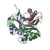
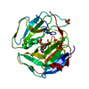
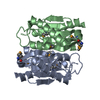
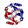
| Title | Crystal Structure of PII from Synechocystis sp. PCC 6803 | ||||||
 Components Components | Nitrogen regulatory protein P-II | ||||||
 Keywords Keywords | SIGNALING PROTEIN / nitrogen regulation / cyanobacteria | ||||||
| Function / homology |  Function and homology information Function and homology informationregulation of nitrogen utilization / enzyme regulator activity / ATP binding / cytosol Similarity search - Function | ||||||
| Biological species |  | ||||||
| Method |  X-RAY DIFFRACTION / MOLECULAR REPLACEMENT / Resolution: 2 Å X-RAY DIFFRACTION / MOLECULAR REPLACEMENT / Resolution: 2 Å | ||||||
 Authors Authors | Xu, Y. / Carr, P.D. / Clancy, P. / Garcia-Dominguez, M. / Forchhammer, K. / Florencio, F. / Tandeau de Marsac, N. / Vasudevan, S.G. / Ollis, D.L. | ||||||
 Citation Citation | Journal: Acta Crystallogr.,Sect.D / Year: 2003 Title: The structures of the PII proteins from the cyanobacteria Synechococcus sp. PCC 7942 and Synechocystis sp. PCC 6803. Authors: Xu, Y. / Carr, P.D. / Clancy, P. / Garcia-Dominguez, M. / Forchhammer, K. / Florencio, F. / Vasudevan, S.G. / Tandeau de Marsac, N. / Ollis, D.L. | ||||||
| History |
|
- Structure visualization
| Structure viewer | Molecule: MolmilJmol/JSmol |
|---|
- Downloads & links
-Download
| PDBx/mmCIF format | 1ul3.cif.gz | 91.7 KB | Display | PDBx/mmCIF format |
|---|---|---|---|---|
| PDB format | pdb1ul3.ent.gz | 69.7 KB | Display | PDB format |
| PDBx/mmJSON format | 1ul3.json.gz | Tree view | PDBx/mmJSON format | |
| Others |  Other downloads Other downloads |
-Validation report
| Arichive directory | https://data.pdbj.org/pub/pdb/validation_reports/ul/1ul3ftp://data.pdbj.org/pub/pdb/validation_reports/ul/1ul3 | HTTPS FTP |
|---|
-Related structure data
| Related structure data | 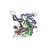 1qy7C 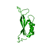 2piiS S: Starting model for refinement C: citing same article ( |
|---|---|
| Similar structure data |


-Links
 PDBj
PDBj- Assembly
| Deposited unit | 
| ||||||||
|---|---|---|---|---|---|---|---|---|---|
| 1 | 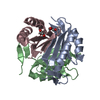
| ||||||||
| 2 | 
| ||||||||
| 3 | 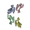
| ||||||||
| Unit cell |
| ||||||||
| Details | chains A, B, C form a trimer / chain D forms a trimer generated by the operations: -y, x-y, z and -x+y, -x, z |
-Components
| #1: Protein | Mass: 12415.374 Da / Num. of mol.: 4 Source method: isolated from a genetically manipulated source Source: (gene. exp.) #2: Chemical |   Mass: 92.094 Da / Num. of mol.: 2 / Source method: obtained synthetically / Formula: C3H8O3 Mass: 92.094 Da / Num. of mol.: 2 / Source method: obtained synthetically / Formula: C3H8O3#3: Chemical |   Mass: 40.078 Da / Num. of mol.: 2 / Source method: obtained synthetically / Formula: Ca Mass: 40.078 Da / Num. of mol.: 2 / Source method: obtained synthetically / Formula: Ca#4: Water | ChemComp-HOH / |  Mass: 18.015 Da / Num. of mol.: 212 / Source method: isolated from a natural source / Formula: H2O Mass: 18.015 Da / Num. of mol.: 212 / Source method: isolated from a natural source / Formula: H2O |
|---|
-Experimental details
-Experiment
| Experiment | Method: X-RAY DIFFRACTION / Number of used crystals: 1 |
|---|
- Sample preparation
| Crystal | Density Matthews: 2.71 Å3/Da / Density % sol: 54.18 % |
|---|---|
| Crystal grow | Temperature: 293 K / Method: vapor diffusion, hanging drop / pH: 4.7 Details: isopropanol, calcium chloride, sodium acetate, pH 4.7, VAPOR DIFFUSION, HANGING DROP, temperature 293K |
-Data collection
| Diffraction | Mean temperature: 293 K |
|---|---|
| Diffraction source | Source: ROTATING ANODE / Type: RIGAKU RU200 / Wavelength: 1.5418 Å |
| Detector | Type: RIGAKU RAXIS II / Detector: IMAGE PLATE / Date: Oct 17, 1997 / Details: monochromator |
| Radiation | Monochromator: GRAPHITE / Protocol: SINGLE WAVELENGTH / Monochromatic (M) / Laue (L): M / Scattering type: x-ray |
| Radiation wavelength | Wavelength: 1.5418 Å / Relative weight: 1 |
| Reflection | Resolution: 2→25 Å / Num. all: 31543 / Num. obs: 30958 / % possible obs: 98.5 % / Observed criterion σ(F): 0 / Observed criterion σ(I): 0 / Redundancy: 4.95 % / Rmerge(I) obs: 0.053 / Net I/σ(I): 16.7 |
| Reflection shell | Resolution: 2→2.1 Å / Redundancy: 2.9 % / Rmerge(I) obs: 0.167 / Mean I/σ(I) obs: 4.93 / Num. unique all: 5151 / % possible all: 98.2 |
- Processing
| Software |
| |||||||||||||||||||||||||
|---|---|---|---|---|---|---|---|---|---|---|---|---|---|---|---|---|---|---|---|---|---|---|---|---|---|---|
| Refinement | Method to determine structure: MOLECULAR REPLACEMENT Starting model: PDB ENTRY 2PII MINUS RESIDUES 37-55 and 109-112 Resolution: 2→25 Å / Isotropic thermal model: overall / Cross valid method: THROUGHOUT / σ(F): 0 / σ(I): 0 / Stereochemistry target values: Engh & Huber
| |||||||||||||||||||||||||
| Displacement parameters | Biso mean: 30.8 Å2
| |||||||||||||||||||||||||
| Refine analyze |
| |||||||||||||||||||||||||
| Refinement step | Cycle: LAST / Resolution: 2→25 Å
| |||||||||||||||||||||||||
| Refine LS restraints |
| |||||||||||||||||||||||||
| LS refinement shell | Resolution: 2→2.02 Å
|
