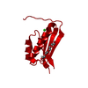
SHEET THE SHEET STRUCTURE OF THIS MOLECULE IS BIFURCATED. IN ORDER TO REPRESENT THIS FEATURE IN ... SHEET THE SHEET STRUCTURE OF THIS MOLECULE IS BIFURCATED. IN ORDER TO REPRESENT THIS FEATURE IN THE SHEET RECORDS BELOW, TWO SHEETS ARE DEFINED.
Mass: 18.015 Da / Num. of mol.: 305 / Source method: isolated from a natural source / Formula: H2O
Compound details
THE SIDECHAIN OF RESIDUE ARG A187 IS OBSERVED IN TWO CONFORMATIONS. A SULFATE MOEITY IS OBSERVED IN ...THE SIDECHAIN OF RESIDUE ARG A187 IS OBSERVED IN TWO CONFORMATIONS. A SULFATE MOEITY IS OBSERVED IN THE VICINITY OF RESIDUE A187, AND HAS BEEN MODELLED TO CO-EXIST ONLY WITH THE A-CONFORMER.
Has protein modification
Y
-
Experimental details
-
Experiment
Experiment
Method: X-RAY DIFFRACTION / Number of used crystals: 1
-
Sample preparation
Crystal
Density Matthews: 1.86 Å3/Da / Density % sol: 50 %
Crystal grow
pH: 8.5 / Details: 100 MM TRIS/HCL PH 8.5, 20% PEG 3350, 200 MM LISO4
Protocol: SINGLE WAVELENGTH / Monochromatic (M) / Laue (L): M / Scattering type: x-ray
Radiation wavelength
Wavelength: 1.07 Å / Relative weight: 1
Reflection
Resolution: 1.5→40 Å / Num. obs: 38985 / % possible obs: 98.1 % / Observed criterion σ(I): 2 / Redundancy: 4.5 % / Rmerge(I) obs: 0.06 / Net I/σ(I): 9.6
Reflection shell
Resolution: 1.5→1.54 Å / Redundancy: 4.5 % / Rmerge(I) obs: 0.27 / Mean I/σ(I) obs: 2.9 / % possible all: 95.4
-
Processing
Software
Name
Version
Classification
REFMAC
5.2.0005
refinement
MOSFLM
datareduction
SCALA
datascaling
SOLVE
phasing
Refinement
Method to determine structure: SAD / Resolution: 1.5→46.47 Å / Cor.coef. Fo:Fc: 0.97 / Cor.coef. Fo:Fc free: 0.955 / SU B: 2.285 / SU ML: 0.04 / Cross valid method: THROUGHOUT / ESU R: 0.087 / ESU R Free: 0.072 / Stereochemistry target values: MAXIMUM LIKELIHOOD / Details: HYDROGENS HAVE BEEN ADDED IN THE RIDING POSITIONS
Rfactor
Num. reflection
% reflection
Selection details
Rfree
0.187
2088
5.1 %
RANDOM
Rwork
0.148
-
-
-
obs
0.15
38985
98.1 %
-
Solvent computation
Ion probe radii: 0.8 Å / Shrinkage radii: 0.8 Å / VDW probe radii: 1.2 Å / Solvent model: MASK
 Movie
Movie Controller


 Open data
 Basic information
 Components
 Keywords
 Function and homology information
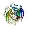
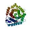
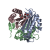
 HUMAN HERPESVIRUS 4 (Epstein-Barr virus)
 X-RAY DIFFRACTION /
  Authors
 Citation
 Structure visualization
 Downloads & links
 Other downloads


 PDBj
PDBj
 Assembly
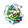


 Mass: 308.182 Da / Num. of mol.: 1 / Source method: obtained synthetically / Formula: C9H13N2O8P
 Mass: 96.063 Da / Num. of mol.: 4 / Source method: obtained synthetically / Formula: SO4
 Mass: 18.015 Da / Num. of mol.: 305 / Source method: isolated from a natural source / Formula: H2O
 Sample preparation
 / Beamline: ID14-4 / Wavelength: 1.07
  Processing