- PDB-1tqc: Ovine recombinant PrP(114-234), ARR variant in complex with the V... -
+
Open data
ID or keywords:
Loading...
-
Basic information
Entry
Database: PDB / ID: 1tqc
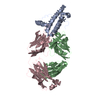
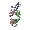
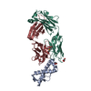
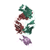
Title
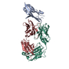
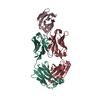
Ovine recombinant PrP(114-234), ARR variant in complex with the VRQ14 Fab fragment (IgG2a)
Components
VRQ14 Fab Heavy chain
VRQ14 Fab light chain
prion protein
Keywords
UNKNOWN FUNCTION/IMMUNE SYSTEM / prion / antibody / UNKNOWN FUNCTION-IMMUNE SYSTEM COMPLEX
Function / homology
Function and homology information
side of membrane / tubulin binding / protein homooligomerization / microtubule binding / copper ion binding / Golgi apparatus / identical protein binding / plasma membrane Similarity search - Function
Prion/Doppel protein, beta-ribbon domain / Major Prion Protein / Prion, copper binding octapeptide repeat / Copper binding octapeptide repeat region / Major prion protein N-terminal domain / Major prion protein bPrPp - N terminal / Prion protein signature 1. / Prion protein signature 2. / Prion protein / Major prion protein ...Prion/Doppel protein, beta-ribbon domain / Major Prion Protein / Prion, copper binding octapeptide repeat / Copper binding octapeptide repeat region / Major prion protein N-terminal domain / Major prion protein bPrPp - N terminal / Prion protein signature 1. / Prion protein signature 2. / Prion protein / Major prion protein / Prion/Doppel protein, beta-ribbon domain / Prion/Doppel beta-ribbon domain superfamily / Prion/Doppel alpha-helical domain / Immunoglobulins / Immunoglobulin-like / Sandwich / Orthogonal Bundle / Mainly Beta / Mainly Alpha Similarity search - Domain/homology
Mass: 12197.569 Da / Num. of mol.: 1 / Fragment: ARR variant, residues 127-228 Source method: isolated from a genetically manipulated source Source: (gene. exp.) Ovis aries (sheep) / Gene: PRNP sheep ARR variant / Plasmid: pET28-a / Species (production host): Escherichia coli / Production host: Escherichia coli BL21(DE3) (bacteria) / Strain (production host): BL21DE3 / References: UniProt: P23907
#2: Antibody
VRQ14FabHeavychain
Mass: 22810.455 Da / Num. of mol.: 1 / Fragment: VRQ14 Fab fragment Source method: isolated from a genetically manipulated source Source: (gene. exp.) Mus musculus (house mouse) / Production host: Escherichia coli (E. coli)
#3: Antibody
VRQ14Fablightchain
Mass: 24155.863 Da / Num. of mol.: 1 / Fragment: VRQ14 Fab fragment Source method: isolated from a genetically manipulated source Source: (gene. exp.) Mus musculus (house mouse) / Production host: Escherichia coli (E. coli)
In the structure databanks used in Yorodumi, some data are registered as the other names, "COVID-19 virus" and "2019-nCoV". Here are the details of the virus and the list of structure data.
Jan 31, 2019. EMDB accession codes are about to change! (news from PDBe EMDB page)
EMDB accession codes are about to change! (news from PDBe EMDB page)
The allocation of 4 digits for EMDB accession codes will soon come to an end. Whilst these codes will remain in use, new EMDB accession codes will include an additional digit and will expand incrementally as the available range of codes is exhausted. The current 4-digit format prefixed with “EMD-” (i.e. EMD-XXXX) will advance to a 5-digit format (i.e. EMD-XXXXX), and so on. It is currently estimated that the 4-digit codes will be depleted around Spring 2019, at which point the 5-digit format will come into force.
The EM Navigator/Yorodumi systems omit the EMD- prefix.
Related info.:Q: What is EMD? / ID/Accession-code notation in Yorodumi/EM Navigator
Yorodumi is a browser for structure data from EMDB, PDB, SASBDB, etc.
This page is also the successor to EM Navigator detail page, and also detail information page/front-end page for Omokage search.
The word "yorodu" (or yorozu) is an old Japanese word meaning "ten thousand". "mi" (miru) is to see.
Related info.:EMDB / PDB / SASBDB / Comparison of 3 databanks / Yorodumi Search / Aug 31, 2016. New EM Navigator & Yorodumi / Yorodumi Papers / Jmol/JSmol / Function and homology information / Changes in new EM Navigator and Yorodumi
 Movie
Movie Controller

 Yorodumi
 Open data
 Basic information
 Components
 Keywords
 Function and homology information
 X-RAY DIFFRACTION /
  Authors
 Citation
 Structure visualization
 Downloads & links
 Other downloads


 PDBj
PDBj


 Assembly

 Mass: 18.015 Da / Num. of mol.: 22 / Source method: isolated from a natural source / Formula: H2O
 Sample preparation
 / Beamline: X06SA / Wavelength: 0.97926 Å
 Processing